How horses go from metabolic to laminitic and ways to manage them
The pasture is particularly verdant this year. Several plump horses contentedly graze their way across the fetlock-deep field in the fog after a particularly cool spring night. This might look like something out of a bucolic period film, but the scene is not as innocuous as it might appear. Several of its features portend potential problems for horses turned out to eat to their hearts’ content. A fat horse on rich grass is at risk of developing endocrine disorders that can snowball into laminitis.
The Facts About Endocrine-Related Laminitis
Laminitis occurs when the laminae (or lamellae)—the tissues that suspend the coffin bone within the hoof capsule—become damaged and inflamed. In severe cases they separate, releasing the coffin bone to rotate downward or sink.
Laminitis is not just localized to the feet, however—it’s also a systemic disease. Only 12% of owner-reported laminitis cases occur due to causes such as colic, diarrhea, retained placenta, or grain overload. The rest are related to diet or obesity and/or systemic endocrine disorders, namely equine metabolic syndrome (EMS) and pituitary pars intermedia dysfunction (PPID, previously referred to as equine Cushing’s disease).
Equine Metabolic Syndrome
Equine metabolic syndrome is not a singular disease but, rather, a compilation of risk factors contributing to endocrinopathic laminitis. Typically, EMS horses are obese with excessive fat deposition and a cresty neck score of at least 3 out of 5. They are predisposed to that weight gain and refractory to weight loss programs. As easy keepers, they tend to develop generalized or regional adiposity (fat pads) if fed too many calories.
Statistics indicate that 51% of mature light-breed horses in the United States are obese. But not all easy keepers on high-calorie diets develop EMS or laminitis. A genetic component might contribute to body condition and alterations in endocrine factors.
Additionally, “thin horses of high-risk breeds can develop endocrinopathic laminitis from other risk factors—horses don’t have to be obese to be susceptible,” says Jane Manfredi, DVM, MS, PhD, Dipl. ACVS-LA, ACVSMR, assistant professor at Michigan State University’s College of Veterinary Medicine, in East Lansing. “Just losing weight isn’t always enough to avoid endocrine issues.”
The nutrition and body condition of a pregnant mare could be another contributing factor to a foal’s endocrine state later in life.
Intestinal health might also play a role. “Less species diversity in the intestinal microbiome is reported in EMS horses, similar to that experienced by humans with metabolic syndrome,” says Manfredi. “Additionally, human metabolic syndrome has been linked with a loss of intestinal barrier function leading to increased intestinal permeability, referred to as ‘leaky gut syndrome,’ which adds to systemic inflammation, which could also be occurring in horses.”
Endocrine-disrupting chemicals (EDCs) are also associated with EMS, with reports that they alter gene expression and affect metabolism. Many EDCs persist in fat for prolonged periods, says Manfredi.

PPID
The other predominant endocrine condition that can lead to laminitis is PPID, which is caused by dysfunction of the pituitary pars intermedia, usually in aged horses. That portion of the pituitary gland overdevelops, leading to excessive production of pituitary hormones, particularly adrenocorticotropin hormone (ACTH), which in turn amplifies cortisol (the stress hormone) secretion from the adrenal glands. Horses with PPID grow shaggy hair and don’t shed normally, their fat is distributed abnormally, and they experience muscle wastage. They tend to drink and urinate excessively and are prone to sinus infections, hoof abscesses, and poor wound healing. Infertility is another common complaint. In 32% of PPID-affected horses, excess circulating insulin (hyperinsulinemia) amplifies the risk of developing laminitis.
Insulin Dysregulation
A consequence of obesity that’s present in many PPID horses is insulin dysregulation (ID), which can further lead to laminitis. Normally, dietary glucose gets absorbed into the bloodstream to stimulate the pancreas to secrete insulin. With ID, cells fail to respond to insulin’s hormonal signals to take up glucose into muscle, liver, and fat cells for storage or metabolism. The pancreas then secretes more and more insulin in an attempt to signal glucose uptake.
Normally, the liver clears nearly three-quarters of secreted insulin. Obese horses and/or those with insulin dysregulation cannot clear it as well.
Adiposity isn’t the only thing that creates insulin overload; intestinal changes contribute, as well. “Specialized endocrine cells present throughout the gastrointestinal tract are distributed along the mucosal surface that lines the gut,” says Manfredi. “Their interaction with intestinal microbiota increases secretion of incretin (stimulating a decrease in blood glucose levels) hormones such as glucagonlike peptides (GLP1 and 2) in response to high nonstructural carbohydrate (NSC) meals, like grain. GLPs are implicated in inducing more pronounced and undesirable insulin responses.”
Cathy McGowan, BVSc, MACVSc, DEIM, Dipl. ECEIM, PhD, FHEA, FRCVS, head of the Department of Equine Clinical Science at the University of Liverpool, in the U.K., has pioneered enlightening research into endocrinopathic laminitis. She describes insulin’s effect on the hoof and, in particular, the lamellae.
“Insulin alters the messages or signaling within lamellar epithelial cells of the hoof, causing them to change their structure—morphing from nice firm little boxes to unstructured blobs with stretched lamellae that won’t spring back,” she explains. “This is due to disruption of the cells’ cytoskeleton (structural shape). Think of a rubber band that loses its spring when left out in the sun. Then imagine, the harder you force that rubber band out of shape (i.e., the more weight put on the hooves), the more it deforms with the potential to break.”
Signs of Endocrinopathic Laminitis
Endocrine-associated laminitis starts with a sometimes-lengthy subclinical (nonapparent) phase that causes chronic changes in the hoof capsule. McGowan says it can develop without warning.
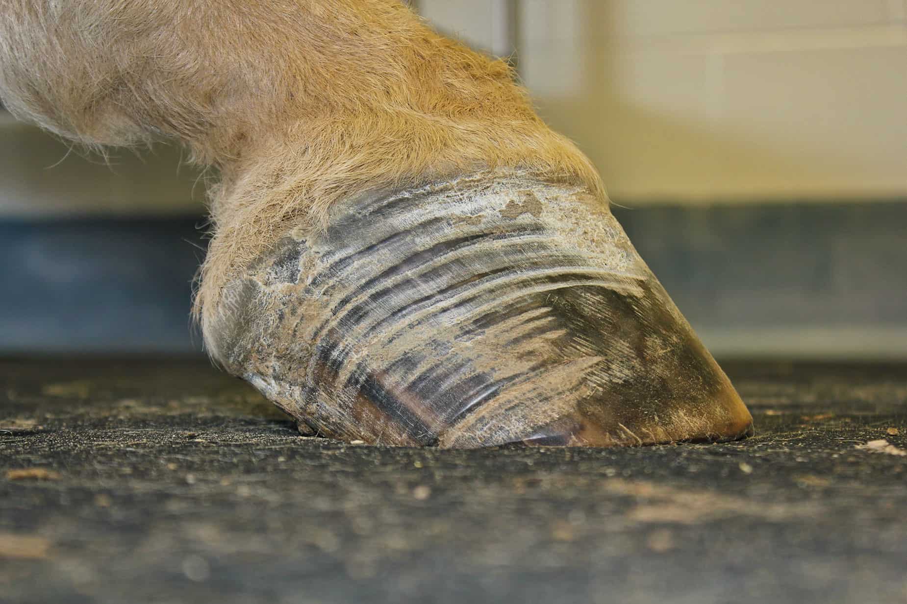
“However, as soon as a horse starts developing telltale divergent hoof rings that are wider at the heel, we know it is a prime candidate for full-blown laminitis any day,” she says. “Some horses can have over a year’s worth of bouts of laminitis with rings extending all the way down the hoof wall, yet have never shown laminitis lameness. Others have more obvious signs, including flaring or distortion of the dorsal hoof wall, a flat or convex sole, and widening of the white line region. These horses are really trying to tell their owners something.”
In other forms of acute and severe laminitis related to systemic inflammatory response syndrome (SIRS) that develops from colitis (inflammation of the colon), endotoxemia (presence of an endotoxin in the blood), uterine infection, or grain overload, inflammatory cells infiltrate the lamellae. The basement membrane that binds lamellar cells together disintegrates, and the lamellae quickly fail. This does not occur with endocrine laminitis, which tends to creep up more slowly, says McGowan.
Armed with the knowledge that most laminitis cases are attributable to systemic endocrine disease, we can implement proactive steps for prevention and treatment. Again, visible changes in the hoof capsule, such as the growth rings, can be evident for months before an affected horse shows lameness. This delay gives owners the opportunity to intervene to control insulin dysfunction. The objective of therapeutic strategies is to prevent stretching and deformation of the lamellae that can ultimately lead to painful laminitis.
Now let’s look at two real-life endocrinopathic laminitis cases to see how the horses’ veterinarians and owners managed them.
1. A Typical Case of EMS Laminitis
An Arabian gelding was actively ridden until the spring of his 17th year, when he suddenly became lame. His owner noticed him being a little “off” over the course of a few weeks and finally acknowledged that her various excuses for why this could be—hard, dry ground; deep snow; uneven terrain; mud—did not add up. She was unclear as to which front leg was lame. Over the course of a week, he became worse—walking stiff-legged and obviously painful. She said it was difficult to pick up a foot for cleaning.
His veterinary exam pointed to clinical laminitis. He had increased (bounding) digital pulses (felt at the back of the fetlock) in both front legs. It was painful for him to turn in a tight circle in either direction. Recent trimming revealed obvious pink areas on his soles, something he’d not had previously. In addition, his body condition score was 7 (on the Henneke scale of 1 to 9), which is significantly overweight. The veterinarian could not feel the gelding’s ribs through the overlying fat layer, his neck had developed a pronounced crest, and he had fat deposits over his shoulders, rump, tailhead, and sheath area. Such fat deposits also generate pro-inflammatory cytokines, which are substances that increase systemic inflammation. He had puffy, swollen areas around his eye orbits, which tend to correspond with PPID.
This horse, whose ideal body weight was 900 pounds, was being fed a disproportionate diet of 18-20 pounds of grass hay a day, plus beet pulp mash and rice bran supplementation. This would be a hefty diet even for a hard-exercising 1,000-pound horse.
The veterinarian had little doubt this horse was suffering from EMS-related laminitis but was also concerned about concurrent PPID, despite his relatively young age. Researchers have found that EMS horses are more at risk of developing PPID, potentially showing signs as early as 10 to 15 years of age. More advanced signs, such as shaggy hair coat and erratic shedding, puffy eyes, topline muscle wasting, and a pot-bellied appearance, take longer to appear despite ongoing hormonal dysregulation. Recurrent laminitis is sometimes the initial presenting sign. Pituitary pars intermedia dysfunction also causes immune suppression and, so, leads to a greater likelihood of developing chronic infections. In addition, cortisol concentrations elevate, which is significant because cortisol decreases insulin sensitivity in the tissues, predisposing these horses to ID and laminitis.
Testing blood ACTH concentrations before and after intravenous administration of thyrotropin-releasing hormone (TRH) is a reliable way to identify early cases of PPID. This horse’s test results confirmed the veterinarian’s suspicions. She immediately started him on pergolide therapy to treat PPID and recommended therapeutic boots as hoof support. She restricted him to 15 pounds of hay (as weighed on a scale) per day, which is within the recommended 1.4-1.7% range of his expected normal body weight for weight loss. She eliminated all other supplements from his diet other than a pelleted dietary balancer (less than 1 pound per day) and free-choice salt. The owner placed his hay in a slow feeder to slow intake and to provide his intestines with small amounts of forage over more time.
“Slow feeders as part of a restricted diet program let foraging time better approximate natural grazing,” says Manfredi. “Offer multiple small meals of hay, ideally three times a day. Once the food is gone, no more is to be fed.”
Prior to feeding, the owner soaked the hay for an hour to remove as much sugar as possible, then poured off and discarded the soaking water.
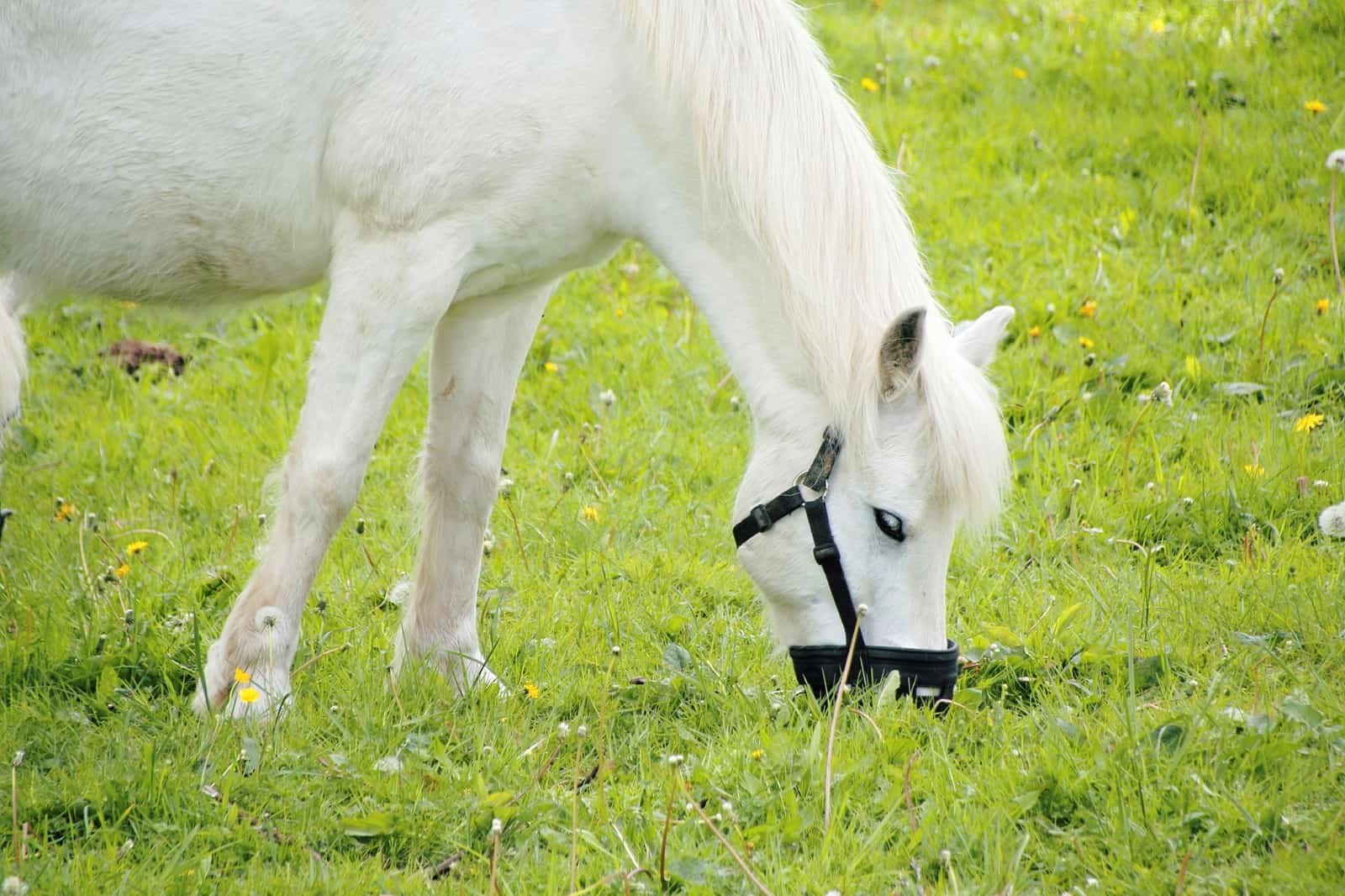
Once his laminitis lameness resolved, the horse was turned out for short periods in the early morning hours, particularly when night temperatures exceeded 40 degrees Fahrenheit. (Grass sugar levels peak in the afternoon and during colder night temperatures.) He wore a grazing muzzle to restrict his intake while still getting light turnout exercise. Grazing muzzles reduce dry matter intake by 77-83%, and exercise helps improve insulin sensitivity.
With these strategies the gelding maintained a good prognosis, because he had no obvious radiographic changes in the position or structure of the coffin bone within the hoof capsule.
While any horse fed a diet rich in calories and NSCs is at risk of developing EMS, Arabians, Morgans, Paso Finos, ponies, Saddlebreds, Warmbloods, and various other breeds tend to be easy keepers with a genetic propensity to lay down fat. These individuals are more at risk for developing EMS than other breeds, and their owners should be proactive about managing weight and body condition.
2. Fighting Obesity and EMS
During a regular spring visit, a veterinarian noticed one patient, a 7-year-old Rocky Mountain horse gelding, was considerably overweight, with a body condition score of 7. Due to her concern that he was developing EMS, she recommended the owner put him on a strict diet, exercise him as much as possible, and only turn him out with a grazing muzzle. About six months later his farrier identified divergent growth rings in his hoof walls and widening of the white line. The gelding soon became overtly lame.
At that time his BCS had elevated to 8, although the owner believed the horse had lost weight. So she had turned him out on green spring grass for four hours a day. The digital pulses in both his front feet had increased. The veterinarian immediately prescribed non-steroidal anti-inflammatories, therapeutic boots, and stall confinement. She advised the owner use a slow-feed haynet with 1.5-by-1.5-inch holes to slow his eating and eliminate insulin surges. She recommended a diet of 16 pounds of soaked hay a day, split into multiple meals in the owner’s net.
On radiographs it was clear the horse’s thin soles were dropped, so he now was relatively flat-footed, adding to his discomfort. He also displayed slight rotation of the coffin bone in both front hooves and thickening of the hoof walls due to scar tissue from chronic inflammatory bouts. The veterinarian talked with the horse’s farrier about improving alignment of the coffin bone and hoof capsule and squaring back the toes to decrease the lever-arm pull of the deep digital flexor tendon on the coffin bone.
The owner kept the horse off pasture and he improved, not experiencing another laminitic bout for two years despite still being overweight. Then, he experienced a recurrence. (A two-year study of 300 laminitis cases found that one-third of horses develop recurrent laminitis, particularly if their insulin elevates and/or they experienced a high lameness grade in their initial diagnosis.)
Testing showed that the horse’s hay was acceptably low in carbohydrates and sugars—ideally, NSC content should be less than 10%. At this point other medication strategies seemed necessary. The veterinarian placed the horse on levothyroxine (a thyroid supplement) for several months to increase his metabolic rate to burn excess energy and improve weight loss. When possible, this strategy works best in conjunction with exercise. (Veterinarians don’t recommend exercise in the painful horse, of course.)
“Given the potential for heart issues secondary to thyroid oversupplementation as shown in recent racehorse research, owners should supplement carefully,” says Manfredi.
The horse also received metformin (an oral antihyperglycemic drug used to treat type-2 diabetes mellitus in humans) 30-60 minutes prior to feeding to reduce glucose absorption from the gut, although Manfredi says results are variable due to poor oral bioavailability (absorption rate). He received vitamin E and an omega-3 fatty acid supplement.
This horse’s resting insulin levels before starting the medication were extremely high; three months later, they were within an acceptable normal range, and his BCS was 6 for the first time in years. Resting insulin is not always the most reliable testing process for predicting ID or laminitis recurrence, but it provided information in this case. McGowan says there is merit in using post-feeding insulin values as a monitoring tool because they more closely approximate the insulin response to a horse’s actual diet. A more labor-intensive evaluation involves dynamic testing with standardized sugar administration to compare insulin and glucose responses to levels before and 60 and 120 minutes after sugar challenge.
In cases like this Manfredi says other possible therapies, including one she’s investigating—a blend of resveratrol and amino acids, including leucine—help decrease inflammation and improve insulin sensitivity. Another medication scientists are currently studying for use in ID horses is velagliflozin (a sodium-glucose-linked transport inhibitor), for its ability to increase glucose excretion through the urinary tract.
Take-Home Message
Seeing your horse every day can make it difficult to recognize body condition changes over time. Controlling obesity is paramount to controlling insulin dysfunction and endocrinopathic laminitis. Take frequent photographs and girth measurements to track weight gain or loss. Learn how to evaluate body condition scores and what’s ideal. And consult your veterinarian about blood tests to monitor your horse’s insulin responses, ACTH concentrations, response to oral sugar challenge tests, and other levels.
Refrain from overfeeding your horse, and especially restrict or eliminate pasture turnout during periods of high sugar accumulation in grass. Your veterinarian can help tailor a dietary program that provides sufficient and safe levels of nutrients compatible with your horse’s needs. Implement lifestyle changes that include not only dietary controls but also regular exercise for your horse.