Afew millimeters deep into the horse’s hoof capsule lies a hoof-shaped bone that seemingly defies gravity. Commonly called the coffin bone, this third phalanx (P3)—analogous to the bone in the tip of the human finger—connects to P2 via a hinge joint. Supported by connective tissues and the sole beneath, this bone, loaded with the horse’s weight in movement, should, in theory, be crashing down with the earth’s pull, causing severe pain.
Amazingly, however, it does not. A remarkable, perhaps superherolike strong matrix of primary fibers called lamellae—and secondary lamellae extending from them like narrow leaves projecting from a book—suspend the coffin bone parallel with the hoof wall. These lamellae grip so strongly onto the coffin bone that they resist even the most powerful, jarring strains placed on them as horses’ feet thunder across the toughest ground.
Laminitis Defined
Laminitis occurs when the lamellae (or laminae)—the Velcro-like tissues that suspend the coffin bone within the hoof capsule—become damaged and inflamed. In severe cases they separate, releasing the coffin bone to rotate downward or sink.
Like all superheroes, though, lamellae have their weak points. Under certain conditions, lamellar tissues weaken, stretch, and separate. Giving way to the load above it, the loosened coffin bone can rotate away from the hoof (tipping downward at the toe) or sink down—even piercing the sole. While insulin appears to be lamellar tissues’ veritable kryptonite, researchers are homing in on other dangers, including abnormal blood flow in the foot and an insufficient energy supply.
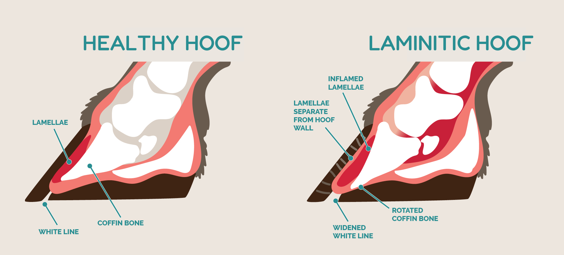
PHOTO: Courtesy Dr. Chris Pollitt
THE 3 CONDITIONS THAT CAUSE LAMINITIS
Scientists have identified three conditions that can provoke laminitis in horses: too much insulin, too much infection, and too much one-sided weight-bearing when the opposite limb is severely lame.
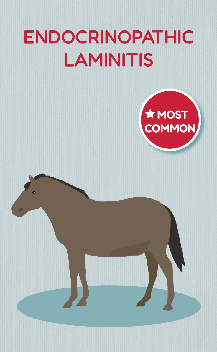
Endocrinopathic, or Metabolic, Laminitis
Insulin is a hormone the pancreas releases to help break down glucose and other sugars; dispensing insulin in normal amounts is a healthy response to sugar. However, insulin-dysregulated individuals release an inappropriate amount of insulin in response to glucose. In humans insulin dysregulation can lead to diabetes. In horses—for reasons we don’t yet clearly understand—it often causes laminitis, says Dr. Chris Pollitt, head of the Australian Equine Laminitis Research Unit at the University of Queensland.
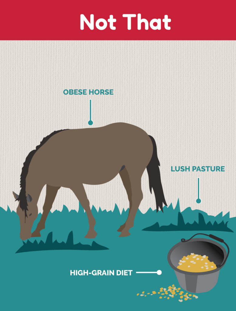
Horses develop insulin dysregulation mainly due to genetics, obesity, aging, and/or pituitary pars intermedia disorder (PPID, also known as equine Cushing’s). Overweight ponies and easy-keeper horse breeds are at the highest risk, but any horse can have insulin dysregulation—even those least likely to have metabolic syndrome, as recent research has shown. “That was an astounding (discovery) to a lot of people, that something like a Standardbred racehorse could get severe laminitis just from having too much insulin in his blood,” Pollitt says.
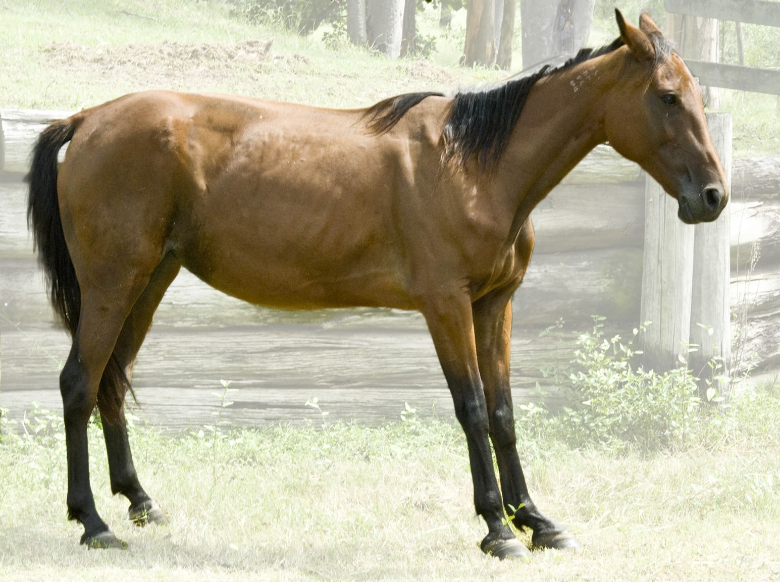
And contrary to popular belief, insulin-dysregulated horses aren’t always fat, says Dr. Andrew van Eps, professor of equine musculoskeletal research at the University of Pennsylvania School of Veterinary Medicine’s New Bolton Center, in Kennett Square. Regular insulin testing is the only reliable way to know which horses are dysregulated and which aren’t. “You can’t just look at a horse and say that horse is healthy,” he states.
Horses can also develop laminitis after insulin spikes from consuming a large amount of sugar—like after breaking into the feed room—or after receiving strong doses of corticosteroids, such as those given in joint injections. “That’s a warning for owners of dressage horses,” Pollitt says. “You give a big shot of a normal dose of corticosteroid, and their insulin’s already elevated? They can go over the cliff.”
Laminitis Caused by Systemic Inflammatory Disease
Infectious diseases such as colitis (infection and inflammation of the colon), placentitis (inflammation of the placenta), pneumonia, sepsis, and Potomac horse fever can also cause certain horses to develop laminitis. While the mechanisms for such laminitis development remain unknown, scientists have speculated it might be due to systemic inflammation and/or the migration of reactive neutrophils (white blood cells capable of engulfing and destroying bacteria and other disease agents, immune complexes, and cell debris) into the lamellar tissue, van Eps says.
Supporting-Limb Laminitis
Finally, horses with severe injuries in one limb can develop laminitis in any of the other three limbs, especially the one opposite the injured leg. Van Eps says this likely occurs because of changes in the normal blood flow pattern in the long-standing foot. Arguably the most famous case of supporting-limb laminitis (SLL) was 3-year-old Thoroughbred Barbaro, who fractured a hind limb in the Preakness Stakes two weeks after winning the 2006 Kentucky Derby. Despite massive veterinary efforts, Barbaro developed severe SLL in his opposing hind limb and had to be euthanized. Study results suggest 12% of all equine patients with a cast show signs of SLL—but that’s likely a significant underestimation, says van Eps.
“It’s the reason we don’t fix a lot of the breakdowns at the track,” he says. “It’s not because we can’t fix the fracture, because with the expertise and modern equipment available, that can be done in many cases without a problem. It’s mostly because they are likely to develop laminitis as a complication.”

Chris Pollitt, BVSc, PhD,
professor emeritus at the University of Queensland, has developed an international reputation as an expert in equine foot biology and disease, in particular laminitis. As head of the university’s Australian Equine Laminitis Research Unit, he has obtained more than $3 million in research funding in the past eight years. Pollitt has contributed 73 publications in international peer-reviewed journals and more than 100 conference proceedings, as well as multiple book chapters. He is the author of The Illustrated Horse Foot: A Comprehensive Guide, published by Elsevier.

Andrew van Eps, BVSc, PhD, MACVSc, Dipl. ACVIM,
professor of equine musculoskeletal research at the University of Pennsylvania’s School of Veterinary Medicine, New Bolton Center, in Kennett Square, is dean of the W. Richardson Endowed Chair of Equine Disease Research provided by the owners of Barbaro, the 2006 Kentucky Derby champion who was euthanized after developing supporting-limb laminitis following a hind-limb fracture. Van Eps devotes his professional life to understanding the mechanisms of all kinds of laminitis—especially supporting-limb laminitis—at a molecular level.


While all three types of laminitis have different modes of action, they consistently lead to the same problem—a mechanical breakdown of the lamellar tissues. “Half the equation is mechanical,” van Eps says. “That’s mostly why we see such disparate, different problems that lead to the same laminitis—your fat hyperinsulinemic horse, your horse with a fracture, your horse with pneumonia. Anything that upsets the cells in the feet or damages them—because they’re under this unique mechanical strain—leads to the same outcome, which is loss of attachment integrity and then pain and the resultant pathology (disease or damage) that we get.”
Because of mechanics, it’s the heavier horses that are at risk of developing the most severe laminitis, he adds. “Pony feet look proportional, but gravity is still a massive factor,” he says. “So for the same amount of lamellar damage a pony might recover quickly, but for a giant Warmblood or Shire it’s going to be a catastrophe.”
PHOTO: The Horse Staff
CLINICIAL SIGNS OF LAMINITIS
In the earliest phases of metabolic laminitis, as the lamellae start to weaken and stretch, horses often show no clinical signs, our sources say. Even with mild inflammation and slight rotation of the coffin bone, many of them—especially leisure and retired horses out at pasture—appear perfectly normal.
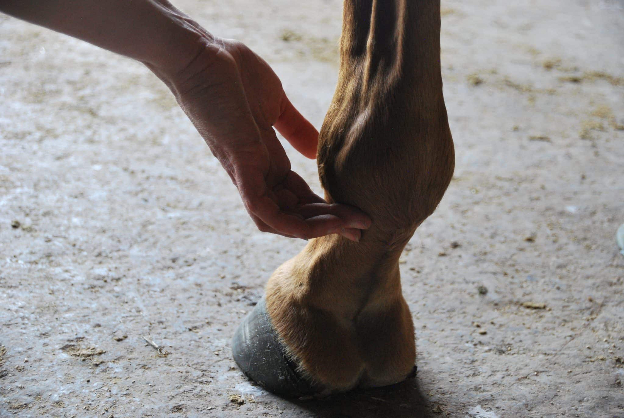
With disease progression, very subtle clinical signs might appear, such as mild lameness when making a turn on hard ground. Regardless of the type of laminitis, horses might also have a strong digital pulse in the artery behind the fetlock, heat in the hooves that lasts for hours, a slight increase in heart rate, or a shortened stride—all signs that van Eps has found reliable across the years. “That hasn’t changed,” he says.
Recently, researchers have discovered that with metabolic laminitis, the higher the insulin levels, the more severe the clinical signs, Pollitt adds. For all types of laminitis, advanced disease causes severe lameness and, as noted earlier, sometimes the coffin bone punctures the sole. Horses often take on a rocked-back stance that shifts hoof loading off the affected toe and toward the back of the foot.
Increasingly, scientists are realizing that poor performance issues in sport horses can also be signs of metabolic laminitis, van Eps says. “Laminitis is not just the crippled pony that can’t walk,” he explains. “We see it as a performance-limiting mild lameness in dressage and some jumping horses.” Eventing horses are less likely candidates because they’re so fit and, so, less likely to have obesity-related metabolic disorders, he adds.
“Laminitis is not just the crippled pony that can’t walk. We see it as a performance-limiting mild lameness in dressage and some jumping horses.”
—Dr. Andrew van Eps
Horses might resist certain movements, fight the bit, swish their tails, or even buck. A sign van Eps says he sees frequently in laminitis cases is the horse that resents getting saddled and girthed by pinning her ears, threatening to bite, and flaring her nostrils. “I’ve seen it many times,” he says. “They know that riding is going to make their feet hurt.”
Poor performance cases are complex because the horses could have laminitis or another orthopedic issue such as navicular disease, or both. Scientists already know humans with metabolic disorders are more likely to have arthritis and other musculoskeletal problems, for example, van Eps explains.
Farriers might see a widening of the white line (between the sole and hoof wall) as the laminae stretch, says Dr. Jaret Pullen, researcher and owner of JP Hoofworks equine podiatry service, in Charlotte, Vermont. But even before that stage, they might notice the horse yanks his feet away when horseshoe nails are driven. “If you have to ease in the nails a little nicer, it starts to make you question the horse’s insulin levels,” he says.
What insulin does to create those signs is a complicated issue, says van Eps. While lamellar stretching is certainly painful, other components—such as nerve damage and neuropathic pain—might be at play “that have nothing to do with the lamellae themselves,” he says.

Jaret Pullen, DVM,
veterinarian, researcher, farrier, and owner of JP Hoofworks equine podiatry service, in Charlotte, Vermont, obtained his veterinary degree from Cornell University. Currently traveling between the northeastern U.S. and Texas, he specializes in treating laminitis, navicular disease, soft tissue injuries, angular limb deformities, and a variety of other complex foot issues. He regularly lectures at equine podiatry seminars and continuing education events in the U.S. and abroad. Pullen is a member of the board of directors for the Northeast Association of Equine Practitioners and actively involved with Colorado State University’s College of Veterinary Medicine’s new podiatry initiative.
PHOTO: Erica Larson
MANAGING AND TREATING LAMINITIS IN HORSES
Laminitis in horses used to be a disease veterinarians handed over to farriers to manage, van Eps says. But today veterinarians are treating the disease from the inside out.
“You’re laboring against a force that you can’t beat,” says van Eps. “You can’t win unless you address the root cause that’s driving the pathology.”
Cryotherapy for Treating Laminitis in Horses
That root cause varies according to whether it’s metabolic, sepsis-related, or supporting-limb laminitis. But one solid across-the-board treatment, regardless of the molecular cause, is cryotherapy. Cooling the horse’s legs inhibits the effects of insulin, controls inflammation, and slows lamellar breakdown and, hence, disease progression, for all three kinds of laminitis, Pollitt explains.
“Our labs gave the profession distal (lower) limb cooling, which meant—especially for the hospital cases with colitis or pneumonia or retained placenta—that the development of laminitis could be arrested, and they could walk out of there and go back to an active life without laminitis,” he says.
In fact, pathological processes drop by 50% for every 10-degree-Celsius temperature drop, Pollitt explains. “So getting a horse foot down from 30 to 5 degrees (86 to 41 degrees Fahrenheit) makes a very big difference. The inflammatory response is stopped in its tracks.” Horses handle the cold temperatures on their legs extremely well, he adds.
Monitoring Insulin Levels in Laminitic Horses
For metabolic laminitis, researchers are taking advantage of their recent discovery of the direct relationship between insulin concentrations and clinical signs, says Pollitt. They now know that as horses’ insulin levels rise, signs worsen—and as concentrations go down, signs improve. That means rapid control of insulin leads to an almost immediate improvement in clinical signs.
“It’s only in the last one or two years that we’ve started to get a real handle on how controlling blood insulin can influence their clinical lameness and even (slow down) radiographic progression,” van Eps says.
“It’s just incredible,” Pollitt adds. “Now, I’m not so upset going to see a case in the field because I’ve got something I can actually do to turn it around.”
Pullen says these findings line up with his field experience. “I was trained mechanically to fix laminitis,” he explains. “But if clients called with a metabolic laminitis case while I was out of town for a week, I’d tell them to work on controlling the insulin—changing diet, soaking hay, drugs if possible—until I got back to do the feet. By then, though, I’d often come back to an almost perfectly sound horse, and that would kind of pop my bubble of all the things I was planning to do to help that horse.”
Hyperinsulinemia
increased insulin—a hormone produced to control blood sugar levels—circulating in the bloodstream 
On the flip side, he says, cases focused on mechanical management alone, without targeting insulin concentrations in horses, were less successful. “No matter what I did, they wouldn’t get better,” he says.
The New Bolton laboratory—among others—can provide insulin test results in one or two hours, van Eps says. And Pullen takes a field-side insulin test on farm calls to get at least a good estimate of a horse’s concentrations. Such fast turnaround times can produce critical information about insulin levels, allowing for immediate treatment. They can also provide frequent, important feedback about how the horse is responding to the treatment in real time.
Drugs Used for Lowering Insulin in Laminitis
Veterinarians have traditionally administered the oral antihyperglycemic drug metformin to help control horses’ insulin levels, but its effects are variable and don’t seem to last more than 10 to 14 days, van Eps says. It also regulates growth-factor signaling and, therefore, might have helpful direct effects on acute laminitis. However, scientists have not yet proven this, he adds.
The diabetes drug pioglitazone can reduce insulin levels through its complex effects on glucose and lipid metabolism, van Eps explains. While veterinarians have used it in horses, they have not established a clear effect in controlling insulin.
Based on scientific studies performed in horses over the past decade, veterinarians have recently turned toward sodium-glucose cotransporter-2 (SGLT2) inhibitors, drugs used commonly in human diabetic patients. They help lower blood glucose by acting on the kidney to increase glucose loss in urine. The reduced blood glucose decreases the stimulus for insulin production in horses, thereby reducing blood insulin levels, explains van Eps.
Ertugliflozin is a popular SLGT2 inhibitor associated with reduced insulin concentrations in insulin-dysregulated horses within days and with “striking improvements in clinical comfort,” says Pollitt, citing work by David Rendle, BVSc, MVM, CertEM(IntMed), Dipl. ECEIM, FRCVS, RCVS, a European specialist in equine internal medicine in the U.K.
Feed and Other Changes for Horses With Laminitis
Diet changes are also critical for reducing insulin levels as well as body weight—which exacerbate laminitis. Horses should be fed a low-carbohydrate forage at 1.5-2% of body weight in dry matter per day and not receive sweet treats, including carrots and apples, Pollitt says.
Meanwhile, the non-steroidal anti-inflammatory phenylbutazone (Bute) can help relieve pain, he says.
Rapid insulin management leads to dramatic improvements in most cases and, despite popular belief, healing of the lamellae. Lamellar tissue is chock full of stem cells and other regenerative components and “actually has a really high capacity for healing,” van Eps says. Granted, it heals in a disorganized fashion, making “an ugly kind of repair” that lacks its original qualities.
A small subset of the horses, however, have endured too much lamellar damage and are simply “too far gone” for effective healing even after insulin levels drop, van Eps says.
The good news is in most cases, if treated early enough, damage and disorganization can be minimal.
“But all it takes is that same metabolic horse to tip over the edge and go back into it again,” Pullen says.
Foot Support and Shoeing for Laminitis Cases
Because mechanics play such an important role in all kinds of laminitis, it might seem useful to get horses off their feet until the lamellae heal, van Eps says. But horses can’t be forced to lie down for long periods, and having no loading on the feet isn’t good for the circulation, either. The goal, then, is to “manipulate the biomechanics of the foot to minimize peak strains.” This provides pain relief while also slowing the disease process, he says.
Farriers can aim for that goal by producing leverage to ease breakover, such as with an omnidirectional shoe or a full rocker shoe, Pullen says. He also generally elevates the heel to reduce tension on the deep digital flexor tendon and supports the back of the foot with frog support to shift the load away from compromised structures.

If the coffin bone has rotated, Pullen says he aims to encourage the new hoof horn growth to be better than the old horn growth, in which “that Velcro attachment (of the lamellae) is gone,” he says. With drugs and good farriery, “you can make that lamellar zone (the distance between the front surface of the hoof capsule and the coffin bone) smaller and tighter, and you can get that bond to be better. There’ll be a section of the hoof wall that’s divergent and has that rotation, but then hopefully the new growth will be parallel as it grows down.”
PHOTO: The Horse Staff/Kevin Thompson
PREVENTING LAMINITIS in horses
Researchers have made significant leaps in treating laminitis in horses over the past two years. Even so, it remains a painful, debilitating disease with irreversible consequences, says van Eps. So, prevention is key. “I think we can’t stress enough how important it is to get on top of the things that are driving laminitis before they actually cause the problem,” he says.
Metabolic laminitis is highly preventable—especially given all we know about it now, van Eps says. If owners can keep their horses’ insulin levels low, their horses won’t develop this form of laminitis. “You have to be very proactive and preemptive and catch these animals early with screening,” he says. “Control the insulin before they get laminitis.”
Regular insulin testing can give a clearer view about how horses’ insulin fluctuates throughout the day, week, month, and season. Horses at risk of insulin dysregulation should undergo insulin testing prior to having a cortisone joint injection to better measure the pros and cons of the procedure, van Eps says.
While drugs can control insulin, smart management is a better long-term goal, he says. “We don’t want to have to medicate animals just so they can live in certain environments.”
Pollitt agrees. “Clients need to have their horses stick to a low-carbohydrate diet, soaked hay, nothing sweet,” he says. “They need to stay in a drylot or have (effective) muzzles on their noses so they can’t eat grass too rapidly.”
People can try planting less glucose-rich pastures, but that’s complicated to do because high-carbohydrate grasses have become dominant in fields, Pollitt says. A more realistic solution is strip grazing—moving an electric or other temporary fence little by little to limit the amount of grass horses can access.
Owners can try track systems such as Paddock Paradise—which also encourage exercise to help keep horses fit—but these can require a big time commitment, Pullen says.
Exercise is actually “one of the biggest components” of controlling insulin, he adds. It’s also one of the trickiest, because horses can’t exercise once laminitis sets in. “They’re fat, they’re sore, they can’t be exercised, and it just extrapolates from there,” Pullen says.
How To Best Cool Horses' Feet
Scientists know cooling horses’ limbs can limit laminitis progression by slowing the biological processes that cause it, even after lameness has developed. It can even prevent the disease if used early enough.
Even so, not all cooling methods are the same, says Dr. Andrew van Eps. To be effective, cooling must act deep enough in the foot to cause the temperature of the lamellae to drop significantly, he says. Van Eps and his colleagues recently embedded temperature probes into the lamellae of five healthy horses. They then tested the cooling effects of four types of commercial cooling boots as the horses stood for four hours, with ice or ice water renewed after two hours. Van Eps presented his team’s findings during the 2023 NAEP Saratoga Vet & Farrier Conference in Saratoga Springs, New York.
Their results showed boots that hold ice in sleeves or via Velcro attachments can lower surface temperatures but not enough to sufficiently cool the lamellae. Boots that keep the horse’s limb constantly immersed in ice water, however, led to effective cooling all the way to the lamellae. Of the two kinds of boots that offer ice water immersion, the one that held the most ice water was most effective, he says. In that boot, lamellar temperatures dropped to as low as 5 degrees C (41 F).
The team also tracked temperatures inside the hoof as six healthy study horses moved freely in stalls for eight-hour periods, wearing the four types of boots, with ice or ice water renewed every two hours. Again, only the two versions that offered ice water immersion successfully kept the foot at low temperatures, with the boot holding the most ice water resulting in the coolest feet, he said.
“Ice water immersion is by far superior, and high-volume ice water immersion is the way to go, at least to about mid-cannon level,” van Eps said.

People—including horse show judges—must recognize what a fit horse looks like and aim for metabolically healthy morphology (form and structure), even in competitions, van Eps adds.
As for sepsis-related laminitis, prevention involves primarily treating the infection and inflammation as aggressively as possible, while keeping all four limbs in cryotherapy boots, which have been shown to reduce the risk of laminitis tenfold in horses with colitis, Pollitt says.
And as for preventing SLL, scientists are still working to find solutions that will keep blood flowing properly in the feet when a horse is recovering from a contralateral limb injury. “We’re now working on trying to improve circulation in horses that aren’t walking or moving around,” van Eps says. That might include new sling technology that loads and reloads intermittently or even forces horses to walk with reduced load, he explains.
PHOTO: Courtesy Penn Vet New Bolton Center
WHAT'S NEW IN LAMINITIS RESEARCH
For metabolic laminitis, researchers are refining methods for detecting endocrine dysfunction and controlling insulin to identify at-risk horses and prevent the problem before it is too late, says van Eps.
Currently, he’s focused on resolving SLL. His team injected cadaver limbs with bloodlike gel in the circulatory system and found the capillaries around the secondary lamellae only filled during a cycle of loading and unloading—as they would with natural stepping, he says.
Positive electron tomography (PET) scanning is providing never-before-seen 3D images of the metabolic health and circulatory system of living horses’ feet. The PET scans, along with other experimental evidence, confirm the importance of cyclic loading (movement) for effective blood perfusion of the feet—including the lamellae. In fact, the blood “chooses” to go through a different path and leaves spaces devoid of blood flow until the foot starts moving normally again, van Eps explains.
“The real kicker, though, is that the damage in horses with fractures is not confined to the supporting limb,” he adds. “I think this reinforces that it’s not necessarily a weight-bearing thing, but it’s a lack of normal load-cycling problem.”
“The real kicker, though, is that the damage in horses with fractures is not confined to the supporting limb. I think this reinforces that it’s not necessarily a weight-bearing thing, but it’s a lack of normal load-cycling problem.”
—Dr. Andrew van Eps
Researchers are realizing horses with sepsis-related laminitis seem to lack glucose and other nutrients in their lamellae, he says. But in these cases it’s not related to poor blood flow. While they’re still trying to find out why that happens, one thing they’ve recognized is icing the foot slows down the lamellae’s entire natural healthy building cycle. In other words, the lamellae don’t need as many nutrients because they’re not actively regenerating their cells. Cryotherapy also appears to control inflammation as well as growth-factor signaling that plays a role in all three forms of laminitis.
“The horse’s foot is a constant source of revelation and new information coming out of research,” Pollitt says. “I’m satisfied that we’ve got a body of work that’s improved things. It’s a journey, and it’s not over yet.”
PHOTO: Getty Images
TAKE-HOME MESSAGE
Metabolic, sepsis-related, and supporting-limb laminitis cause mechanical failure of one of the horse’s most impressive biomechanical designs: the suspension of the last toe bone inside the hoof by a lamellar network. Laminitis can destroy a horse’s athletic career and even be fatal, but researchers are making significant advances in understanding this complex disease while finding effective treatments. More importantly, by focusing on insulin testing and control, cryotherapy, and circulation in the foot, they’re finding methods that might one day prevent the irreversible damage of laminitis altogether.
Credits
 Christa Lesté-Lasserre, MA
Passionate about horses and science from the time she was riding her first Shetland Pony in Texas, Christa Lesté-Lasserre writes about scientific research that contributes to a better understanding of all equids. After undergrad studies in science, journalism, and literature, she received a master’s degree in creative writing. Now based in France, she aims to present the most fascinating aspect of equine science: the story it creates. Follow Lesté-Lasserre on Twitter @christalestelas.
Editorial Director: Stephanie L. Church
Managing Editor: Alexandra Beckstett
Digital Editor: Haylie Kerstetter
Art Director: Claudia Summers
Web Producer: Jennifer Whittle
Publisher: Marla Bickel