


Management factors such as the amount, type, and timing of roughage provided, the amount of non-structural carbohydrates per meal, and the amount and timing of exercise can disrupt these normal protective mechanisms and cause acid to splash up on the squamous mucosa. Transport is also a recognized risk factor.
Roughage
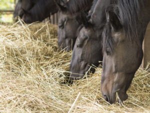
Adequate roughage is the cornerstone of good gastric health, as well as being one of the 3 Fs (Friends, Forage, & Freedom) essential for good equine well-being. The amount, type, and timing of roughage all influence its ability to protect the stomach.
A minimum of 1.5% bodyweight/day DM of hay is commonly recommended. However, this recommendation is based on dry matter intake, and that all roughage, including hay, includes some water. As such, an easier and more appropriate recommendation is the provision of at least 2% bodyweight/day of a suitable hay as fed. This is approximately 20-25 pounds of hay for a typical 1000–1250-pound horse, each day. Importantly, the definition of what a suitable hay is varies between horses, depending on factors such as their underlying metabolic state and level of exercise, and it is important to match the type of hay to each horse’s needs. In some cases, this means intentionally selecting a lower nutritional quality hay to ensure the metabolic needs of the horse is met while still aiming for a 2% bodyweight/day as fed target.
The type and physical structure of the hay also influence the amount of chewing required to eat. Chewing is essential for gastric health because each chewing cycle produces saliva, which contains bicarbonate as a natural buffer against gastric acid. As such, long-stem, stalkier hay is preferred because it encourages more chewing as well as more effectively forming the roughage ball needed to protect the stomach. Alfalfa has some beneficial properties, over and above grass hay, as it has additional buffering capacity. However, the benefits of this are modest in comparison to the overall benefits of consuming an appropriate amount of hay each day, and ensuring intake of an appropriate amount of roughage should be the first priority in ration formulation.
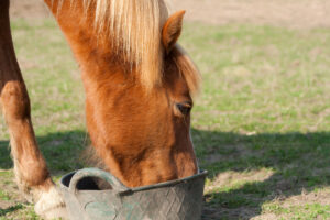
Pasture turnout is another of the essential 3Fs (Freedom), and has numerous benefits for horses, including increasing the amount of time spent eating. However, pasture turnout itself is not automatically protective against squamous disease, as the length, type, and amount of pasture available all influence chewing behavior and the ability to form a protective roughage ball in the stomach. In many management conditions, the provision of hay in addition to pasture turnout is optimal for gastric health.
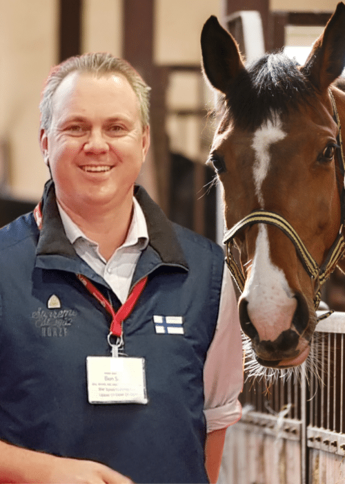
Giving free-choice hay does not automatically translate into improved gastric health. Eating hay is a complex behavior, and horses must feel safe and secure to eat normally. As such, addressing the overall housing of horses with a history of squamous disease can have large, positive impacts on eating behavior. For example, horses housed outdoors spend nearly 50% more time eating than horses housed indoors, and horses housed with other horses spend nearly 40% more time eating than horses housed in isolation. This increased time spent eating translates into greater roughage consumption, maintenance of the roughage ball, and greater saliva and bicarbonate production. “Weighing and measuring individual hay intake is a great first step in understanding why an individual horse might have squamous disease, even if it is provided with free-choice hay,” says Sykes. “It’s remarkable how common horses with access to unlimited good quality hay are still consuming significantly less than the target 2% bodyweight/day as fed recommendation. This is because eating is a complex behavior, and when we identify this, it points us to look at the broader aspects of the horse’s environment and their impacts on behavior, as well as considering other potential contributors such as dental disease.”
Carbohydrates
Excessive non-structural carbohydrates (NSCs) have been shown to increase the risk of squamous disease, but NSCs are not inherently bad, and they do not need to be completely avoided. In fact, appropriate amounts of NSCs are important for optimal performance in many of our athletic horses. So, instead of trying to remove NSCs from the diet completely, the recommended approach is to ensure an appropriate amount is fed each meal, with 1 g/kg/meal the upper limit recommended. The adoption of modern, “low carb” diets in U.S. sports and recreational horses means that many horses are safely within the limits under current management conditions. In other usage types, such as high-level eventers, racehorses and endurance horses, higher energy demands mean that NSC loads in diets tend to increase. Rigorously evaluating diets and considering simple strategies such as including oil in the diet or increasing the number of meals/day (smaller meals, more frequently) to maintain the NSC load below 1 g/kg/meal is useful in these populations.
Balancing the relative benefits of different types of hay and NSC load while meeting the overall nutritional needs of any individual horse can be challenging, and consulting your veterinarian or trained equine nutritionist can be beneficial in meeting the competing demands of diet.
Exercise
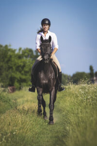
Exercise plays a key role in the risk of disease, specifically the duration of exercise at a trot or above. During exercise, acid is pushed up from the ventral part of the stomach onto the dorsal, squamous part of the stomach. Squamous ulcers are effectively an acid burn, so the longer this exposure occurs, the more likely a burn (i.e. ulcer) will occur and the worse the burn (ulcer) will be. The exact amount of exercise at which squamous risk increases is not well defined, but current recommendations are to, wherever possible, keep the cumulative duration of exercise at a trot or above to less than a total of 30-40 minutes per day. This is achievable in many recreational and riding horses, often by stepping down to a walk during the cool-down period sooner in the exercise cycle but can be challenging in horses with higher inherent workload requirements (e.g. endurance horses and eventers). In these populations, recognizing the increased risk and adopting additional strategies (such as targeted use of appropriate supplements or omeprazole) warrants consideration.
Timing of exercise is another important factor. Horses naturally fast overnight, reducing their roughage intake in favor of resting. If they are exercised first thing in the morning, before an adequate amount of roughage has been consumed, it means that no roughage ball is present in the stomach, resulting in extensive acid splash (image 2). Recognizing this as a risk factor is important in certain populations (e.g. Thoroughbred racehorses that typically work early in the morning) or under certain management conditions (e.g. early morning riding to avoid heat later in the day). When recognized as a risk factor, the addition of pre-exercise feeding, ideally with long-stem alfalfa hay, is a useful intervention to reduce squamous disease risk. Where pre-exercise feeding is not possible, often due to horses having limited appetite in the early morning (e.g. Thoroughbred racehorses), adopting additional strategies (such as targeted use of appropriate supplements or omeprazole) could be added.

 Stacey Oke, MS, DVM, is a practicing veterinarian and freelance medical writer and editor. She is interested in both large and small animals, as well as complementary and alternative medicine. Since 2005 she’s worked as a research consultant for nutritional supplement companies, assisted physicians and veterinarians in publishing research articles and textbooks, and written for a number of educational magazines and websites.