
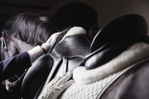
Lameness in the Gaited Horse
- Topics: Article
Approximately 40 participants gathered at the Table Topic discussion on Lameness in the Gaited Horse, which took place at the 2008 American Association for Equine Practitioners convention, held Dec. 6-10 in San Diego, Calif. Facilitators opened the floor with a brief explanation of the AAEP Tennessee Walking Horse Task Force's white paper, Putting the Horse First: Veterinary Recommendations for Ending the Soring of the Tennessee Walking Horse. Monty McInturff, DVM, of Tennessee Equine Hospital in Thompsons Station, Tenn., explained as members of the AAEP, attendees should all stand behind this work that will be the model for elimination of soring in the Tennessee Walking Horse. There were comments from practitioners representing both the Morgan and American Saddlebred Horse breeds, stating that they had heard many positive breed industry comments in support of this white paper. Everyone in the room showed support for maintaining the welfare of all gaited breeds.
The next topic of discussion focused on stifle lameness. John Bennet, DVM, of Bell Buckle, Tenn., explained the presentation of this lameness, which can be as subtle as a momentary patellar fixation (hitching) or a full patellar lock. He stated that this is most commonly seen in young horses without proper muscle development or animals with some underlying condition. The first diagnostic step is to determine if the stifle is radiographically normal (showing no signs of osteochondrosis dissecans or degenerative joint disease). If it is normal, the options are physical therapy with regular work schedules, injections to stimulate the patellar ligament, or surgical intervention.
The two surgical techniques discussed were patellar ligament fenestration and patellar ligament desmotomy. Fenestration can be performed on the standing horse with sedation and a local analgesic. Veterinarians can perform the procedure with a 16-gauge needle or a #15 scalpel blade, making multiple stab incisions along the full length of the medial patellar ligament. The desmotomy is also performed with standing sedation and a local cutting of the medial patellar ligament at the level of its insertion
Create a free account with TheHorse.com to view this content.
TheHorse.com is home to thousands of free articles about horse health care. In order to access some of our exclusive free content, you must be signed into TheHorse.com.
Start your free account today!
Already have an account?
and continue reading.
Related Articles


Stay on top of the most recent Horse Health news with


