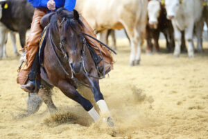
The DFTS is more than just the synovial casing that surrounds the flexor tendons as they course around the bottom third of the cannon bone and the fetlock joint and disperse throughout the pastern region, said Florent David, DVM, MS, Dipl. ACVS & ECVS, Dipl. ACVSMR, ECVDI Assoc., specialist in Surgery, Sports Medicine & Rehabilitation, and Diagnostic Imaging at the Equine Veterinary Medical Center, in Doha, Qatar. He reviewed the anatomy and described how he diagnoses and treats closed DFTS injuries at the 2019 Northeast Association of Equine Practitioners Symposium, held Sept. 25-27 in Saratoga Springs, New York.
DFTS Anatomy
The three continuous, communicating (shared fluid space) regions throughout the length of the sheath meet several bony prominences along the way, where the superficial digital flexor tendon (SDFT) and the deep digital flexor tendon (DDFT) encounter considerable changes in direction and friction.
Other parts—mesotenons, thin stabilizers connecting the tendons to the sheath and providing nutrients and oxygen to the tendon via blood vessels, and tendinous rings called manica flexoria or annular ligaments, for instance—help with the intricate mechanics. Some of these parts are difficult to see on imaging except in horses with DFTS inflammation
Create a free account with TheHorse.com to view this content.
TheHorse.com is home to thousands of free articles about horse health care. In order to access some of our exclusive free content, you must be signed into TheHorse.com.
Start your free account today!
Already have an account?
and continue reading.