Ultrasound for Arytenoid Chondritis Diagnosis?
Veterinarians often choose upper airway endoscopy when working to diagnose equine arytenoid chondritis–an uncommon but problematic respiratory condition–but in some cases a definitive diagnosis lies out of reach. Ultrasonography could offer a valuable adjunct tool for diagnosing this respiratory condition, however, especially in cases lacking a definitive diagnosis.
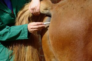
At the 2012 American Association of Equine Practitioners convention, held Dec. 1-5 in Anaheim, Calif., Katherine Garrett, DVM, Dipl. ACVS, of Rood & Riddle Equine Hospital, in Lexington, Ky., presented results of a study in which she and colleagues evaluated ultrasonographic features of horses with arytenoid chondritis.
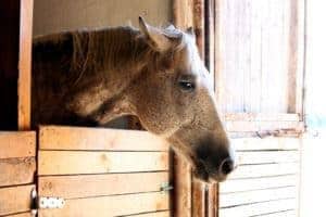
Arytenoid chondritis is inflammation involving one or both arytenoid cartilages, which close over the opening to the trachea when a horse swallows. Affected performance horses often exhibit diminished performance, cough when exerted, and make abnormal noise during exercise (similar to "roaring").
Garrett noted that while ultrasonography has potential to help veterinarians diagnose arytenoid chondritis, specific ultrasonographic parameters had not been identified. This led Garrett and colleagues to compare ultrasonographic findings of horses with confirmed arytenoid chondritis (diagnosed via endoscopy) and horses with normal arytenoid cartilage function and structure
Create a free account with TheHorse.com to view this content.
TheHorse.com is home to thousands of free articles about horse health care. In order to access some of our exclusive free content, you must be signed into TheHorse.com.
Start your free account today!
Already have an account?
and continue reading.

Related Articles

Stay on top of the most recent Horse Health news with