A New Navicular Vantage
Technological advances such as MRI have given veterinarians a closer look at navicular syndrome.

Technological advances such as MRI have given veterinarians a closer look at navicular syndrome.
A mobile MRI suite will be available at Helen Woodward Equine Hospital in Rancho Santa Fe, Calif., as needed.

MRIs picked up a wider range of abnormalities in some lame horses than did X rays or ultrasounds.
Virginia Tech’s Marion duPont Scott Equine Medical Center (EMC) has announced that they are now able to offer high field MRI service for horses. A mobile high field MRI is scheduled to the clinic in Leesburg, Va., about once a month, according to a

Dr. Katherine Garrett of Rood and Riddle Equine Hospital in Lexington, Ky., discusses the use of MRI to diagnose injuries and other problems in horses.
Pinpointing lameness in horses is crucial before proper treatment can be prescribed. There are several approaches to lameness examinations and diagnostic methods, which will be partly determined by the age of the horse and what it does for living.
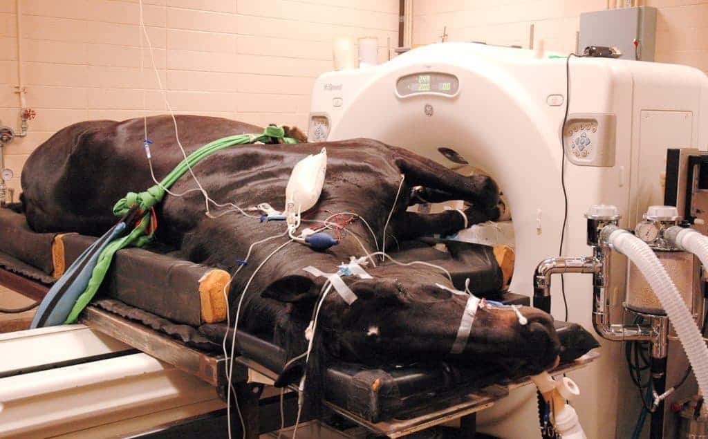
A new clinical imaging system in place at the University of Florida (UF) Veterinary Medical Center will enable veterinarians to obtain diagnostic images of previously inaccessible and larger parts of the body, such as the upper legs of horses, veterinarians say.
The LSU School of Veterinary Medicine recently acquired a Hitachi Echelon 1.5 Tesla MRI unit. This is the first and only high field MRI unit in the state of Louisiana for veterinary use.
In earlier years, a diagnosis of navicular disease was often considered career-ending for a horse. Chronic lameness was typical, in spite of therapeutic
The high degree of detail seen with MRI has made it possible for veterinarians to find equine injuries they’ve never seen before. One example of this–MRI evaluation of desmitis in the oblique and straight distal sesamoidean ligaments–was discussed.
Not all MRI units are created equal. Learn about the differences in MRI units.
Pain originating in the upper cannon bone area, just below the knee or hock, is common in all types of equine athletes. However, it can be difficult to determine exactly what structure is injured; some injuries can only be seen with high-field MRI.
For decades veterinarians have relied upon a number of different imaging tools, from radiographs to ultrasonography, to diagnose lameness, pregnancy, and
Veterinarians now have more tools to help diagnose and treat specific lameness problems.
Take more than 1,000 veterinarians and veterinary students from around the globe, some armed with presentations representing thousands of hours worth of equine research, and add to it some tartan, bagpipes, and Scottish fare. Drop it all into a
Each year at the British Equine Veterinary Association Congress (BEVA), the organization awards prizes to top student presentations in the clinical research portion of the program. The 2007 award winners? talks were both rooted in orthopedics,


