Non-Weight-Bearing Ultrasound
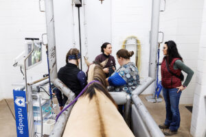
Veterinarians can take non-weight-bearing ultrasounds of horses’ lower limbs with the foot elevated in a Hickman block (a hoof positioning block used for imaging), with someone holding the limb in a flexed position, or with the limb (usually a hind limb) resting on its toe. Most horses require sedation to remain still during these procedures.
Fairburn said veterinarians choose to perform non-weight-bearing ultrasound for multiple reasons:
- It improves the ultrasound probe’s contact with the limb due to laxity (looseness) of the superficial digital flexor and deep digital flexor tendons that run down the horse’s leg. “You can just kind of squish them and get a much broader contact area,” she explained.
- It reduces tension on the structure of interest.
- Because of the latter two effects, lesions might become more obvious. “Lesions may increase in size, and marginal tears may open up,” she said.
Off-Incidence Ultrasound
Veterinarians can perform off-incidence ultrasound (when the beam is not at 90 degrees) with the horse weight-bearing or non-weight-bearing. The technique involves placing the probe over the affected area at a normal angle, or incidence, then dropping the tail of the probe by a few degrees until the tendon or ligament goes black on the screen, Fairburn explained.
“It’s useful for delineating the margins of structures and identifying chronic injuries, because scar tissue will remain white when you go off-incidence,” she said.
Doppler Ultrasound
Veterinarians have two types of Doppler ultrasound at their disposal: color and power. They both offer information about the speed and direction of blood flow, with power Doppler providing greater detail.
“Broadly, they’re just telling us about blood flow in tendon and ligament tissue,” said Fairburn, which can help veterinarians identify areas of pathology and assess healing over time. “Hypervascularity occurs in the normal healing process, so we see an increase in blood flow that tails off as healing progresses. It normally lasts three to six months after injury.”
While normal, healthy tissue doesn’t typically show blood flow, Fairburn cautioned that you can detect it immediately after exercise. “So if I’m thinking about using Doppler, I’ll wait a while after the horse exercises.”
Veterinarians should perform Doppler ultrasound in non-weight-bearing limbs and optimize the machine for low-flow velocity, she said, adding that they should take care not to press so hard on the tissue that they compress the blood flow themselves, she added.
Dynamic Ultrasound
This procedure, also done non-weight-bearing, allows veterinarians to examine a region of the limb during flexion and extension and assess relative movements of the structures. Fairburn said she most commonly performs dynamic ultrasound when imaging the digital flexor tendon sheath.
Take-Home Message
Non-weight-bearing and dynamic ultrasound can provide practitioners with greater details about lesions they’ve already identified and allow them to identify lesions they haven’t identified previously. Fairburn said these techniques are relatively easy to do and require no extra equipment compared to weight-bearing ultrasound—just practice.