Diseases Affecting the Equine Vertebral Column

Equine submissions to the University of Kentucky Veterinary Diagnostic Laboratory over a 10-year period (2011-2021) were queried for diagnoses related to significant vertebral column pathology. A total of 426 cases were identified. Distribution of diseases included equine cervical vertebral stenotic myelopathy (n=224), fracture/subluxation (n=123), abnormal spinal curvature (n=51), osteomyelitis (n=13), intervertebral disc disease (n=7), congenital vertebral anomaly (n=5), and neoplasia (n=3).
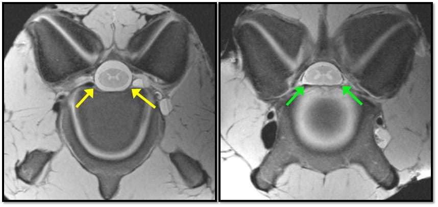
Vertebral pathology associated with equine cervical vertebral stenotic myelopathy (wobbler syndrome) comprised the majority of cases. This is no surprise given the density of Thoroughbreds in the central Kentucky area and their documented breed predisposition to develop the disease. Wobbler syndrome occurs when malformation of the cervical vertebrae results in spinal canal narrowing and cervical spinal cord compression. In general, wobbler syndrome is categorized as affecting two groups of horses. The first being young, growing horses with complex multifactorial interactions between gender, growth rate, diet, and genetic determinants. The second group is seen in older horses with age-related changes in the neck, mainly osteoarthritis. In this retrospective, males were more commonly affected (male n=182; female n=34), the average age was 23.1 months (4-168 months), and Thoroughbreds were the most common breed (n=165). Vertebrae are classified as irregular bones due to their complex shape. Therefore, changes in different locations of the bone itself can result in spinal cord compression. Articular process joint lesions were the most frequent cause of spinal cord compression (n=71) followed by subluxation of the vertebral body (n=67), generalized narrowing of the canal (n=27), and thickening or elongation of the dorsal lamina (n=13). Articular process lesions were largely due to osteoarthritis, followed by osteochondrosis.
The cervical column was the most commonly reported site of fracture/subluxation (n=69), with the vertebral body being most frequently affected. In 11 cases, partial spinal cord transection was described. While a clinical history of trauma was reported in all identified cases, six cases had evidence of underlying neurologic disease (wobbler syndrome or equine protozoal myeloencephalitis).
Acquired spinal curvature disorders (i.e., scoliosis, lordosis, or kyphosis) were identified in 51 cases, with 50 considered congenital and one case of acquired scoliosis. Of the congenital cases, 71% had other skeletal abnormalities, including limb contracture or facial malformations. Dystocia was described in 66% of the submitted histories. The thoracic column was the most common location (n=27), followed by the lumbar column.
All vertebral osteomyelitis cases occurred in horses under one year of age. The lumbar vertebrae were the most frequent location. Rhodococcus equi and Streptococcus zooepidemicus were common bacterial isolates. Secondary pathologic fracture was noted in six cases.
Clinically significant intervertebral disc disease is a more recent area of interest. In this review, intervertebral disc disease cases exhibited yellowing of the disc material with varying degrees of fibrillation, clefting, and loss. All cases had a clinical history of ataxia. Secondary spinal cord compression was noted in four cases.
Of the remaining disease categories, congenital vertebral anomalies included cystic vertebral body or vertebral fusion. Neoplastic processes impacting the vertebral column included primary vertebral body sarcoma, metastatic lymphosarcoma, and metastatic melanoma.
A variety of disease processes can impact the equine vertebral column. Pathologic evaluation of the equine vertebral column in conjunction with clinical information will continue to help better our understanding of these processes and their impact on equine health.
Editor’s note: This is an excerpt from the University of Kentucky’s Equine Science Review, Issue 21, published on March 5, 2022. Jennifer Janes, DVM, PhD, Dipl. ACVP, assistant professor of anatomic pathology at the University of Kentucky Veterinary Diagnostic Lab, provided this information. Source: January 2022 Equine Disease Quarterly.
Written by:
University of Kentucky College of Agriculture, Food, and Environment
Related Articles
Stay on top of the most recent Horse Health news with


