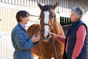
Oskie, a 20-year-old Arabian gelding, had numerous mild colic episodes over the past two years. His owner is a veterinarian and knew how to treat the increasingly chronic condition in Oskie. But when she treated him a dozen times in one month, it was time to have his condition more thoroughly examined by equine specialists at the University of California, Davis, veterinary hospital.

She suspected Oskie might have enteroliths, which are rocklike formations that form in the intestinal tract and cause blockage. Consisting of sand and other undigestible items that collect in the colon, enteroliths range in size from little pebbles to bowling balls if allowed to grow long enough.
At UC Davis’ Large Animal Clinic, Oskie underwent a series of examinations in several departments, which began with a thorough work-up by faculty member Dr. Julie Dechant, DVM, MS, Dipl. ACVS, ACVECC, and resident Stefanie Arndt, DVM, DrMedVet, of the Equine Emergency Surgery Service. Radiology specialists with the Diagnostic Imaging Service detected no enteroliths or sand in his system on X rays, so he was referred to the Large Animal Ultrasound Service for an abdominal ultrasound. There, Betsy Vaughan, DVM, Dipl. ACVSMR, observed a long segment of severely distended and fluid-filled small intestine that had poor motility. This appearance was highly suggestive of a small intestinal obstruction, though the cause of the obstruction could not be seen, so Vaughan recommended surgery to find the cause.

Surgeons performed an exploratory celiotomy, an opening of the abdominal cavity, and found a mass growth blocking Oskie’s small intestine and removed a 4-foot section of the intestinal tract. The appearance of the mass was consistent with cancer, but there was no visual evidence of it spreading to the adjacent intestine or lymph nodes.
A biopsy of the mass by the Anatomic Pathology Service confirmed it to be a jejunal adenocarcinoma, a rarely found cancer in horses. With large margins surrounding the tumor removed and no metastasis observed in his system, Oskie appeared free of the cancer following surgery.
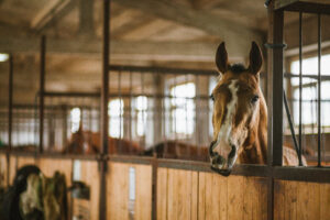
Oskie was weak following surgery, but sling assistance allowed for a smooth recovery. He was hospitalized in the Equine Intensive Care Unit for 10 days while being treated with intravenous fluid therapy, antibiotics, non-steroidal anti-inflammatory drugs, and nutritional support.

Horses that undergo colic and other abdominal surgeries have long recoveries. For the first 30 days, Oskie was confined to stall rest with only two short hand walks per day. For the second month, he was allowed access to a small run along with his hand walks. The third month of Oskie’s recovery gave him access to a large pasture by himself and continued hand walks.
At Oskie’s three-month recheck appointment, a follow-up ultrasound showed his small intestine was back to normal compared to its previous dilated appearance. Now 10 months post-surgery, his owner reports he has made a full recovery.