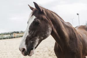
A regularly scheduled veterinary oral examination is the foundation for good oral hygiene. Veterinarians emphasize preventive routine oral care through all life stages, from the juvenile to the geriatric patient. The basis of general dentistry in horses is to encourage the teeth to wear down evenly and thereby wear away slowly. Our goal is to preserve and promote functional dentition for as long as possible. The maintenance of good oral health and comfort fosters increased overall value, allowing our patients to lead long, useful, and well-cared for lives.
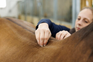
A routine, lightly sedated oral examination is at the core of modern general dentistry. We recommend every horse have an oral consultation performed at least once a year. Occasionally, more frequent examination and care is recommended. During every visit veterinarians perform a thorough oral and dental evaluation to assess external structures and head symmetry, dental occlusion, oral soft tissues, and endodontic (soft tissue inside the tooth) and periodontal (structures that support the tooth) health.
Following each veterinary oral exam, sharp points and dental overgrowths are carefully reduced only if necessary. The significance of any identified pathology is reviewed, and further diagnostic and treatment options are discussed. Dental X rays might be recommended to get a more complete oral health picture. At every appointment, patient comfort and owner education are a priority.
Periodontal Disease in Horses
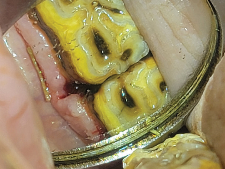
Gum disease, known by veterinary dentists as periodontitis, is a very painful condition in horses. Researchers have suggested that up to 40% of horses are affected. What starts as inflammation of the gums around affected teeth, if left unmanaged, spreads beneath the gum line and leads to infection of the bone that holds the teeth in place. Advanced cases of periodontal disease result in bone loss and mobile teeth, with potential infection of the sinuses or jaw bone and an end result of tooth loss.
In horses, gum disease is usually an orthodontic issue caused by teeth that are misplaced. Orthodontic misalignment results in abnormal spaces between teeth, which veterinary dentists refer to as diastema. Food entrapment in these spaces initiates the gum inflammation and, later, periodontal disease. If identified early, the inflammation can be halted or sometimes reversed. When unmanaged or diagnosed in later stages, permanent soft tissue and bone loss might have already occurred.
Our goal is always to increase oral comfort and halt or reverse disease progression when possible. We use a variety of periodontic management options available and treat each case individually, taking into consideration the patient’s age, breed, history, and stage of periodontal disease.
Teeth Extractions in Horses
Some basic extractions can reasonably be performed in the field such as wolf teeth, retained caps, or loose teeth with more than 3 millimeters of mobility. However, most other extractions are completed at the clinic for the safety and comfort of the patient and to deliver the necessary resources for success.
We perform most all extractions in the standing horse with good head support, deep sedation, and regional and local nerve blocks to provide a comfortable and pain-free patient experience. Preoperative bloodwork, which might include a complete blood count and chemistry profile, is usually performed prior to the procedure to ensure the patient does not have underlying health concerns.
Extractions are indicated mostly due to crown fractures, tooth root infection or resorption, or the presence of advanced periodontal disease. For diseased incisors, simple oral extraction can sometimes be performed. For diseased canine teeth, which have a long, curved root fixed into the jaw, a surgical approach requiring a flap and bone removal is typically necessary.
Simple intraoral extraction is always the preferred method of cheek tooth extraction and is the technique we first apply when applicable. Simple extraction carries the least risk of complication but is only possible when there is sufficient crown stable enough to withstand the force of extraction forceps.
For complicated extractions, when simple extraction is not possible, we can perform advanced standing surgical techniques. We transition to surgical extraction techniques most frequently to extract decayed teeth with brittle crowns or crowns that have previously fractured or to remove malformed teeth, or when the socket is not wide enough for simple extraction of the tooth. In some cases, the use of multiple techniques is necessary. Pre- and postoperative X rays are routinely performed as part of an extraction.