Get a clearer picture of what happens when a horse’s immune system malfunctions
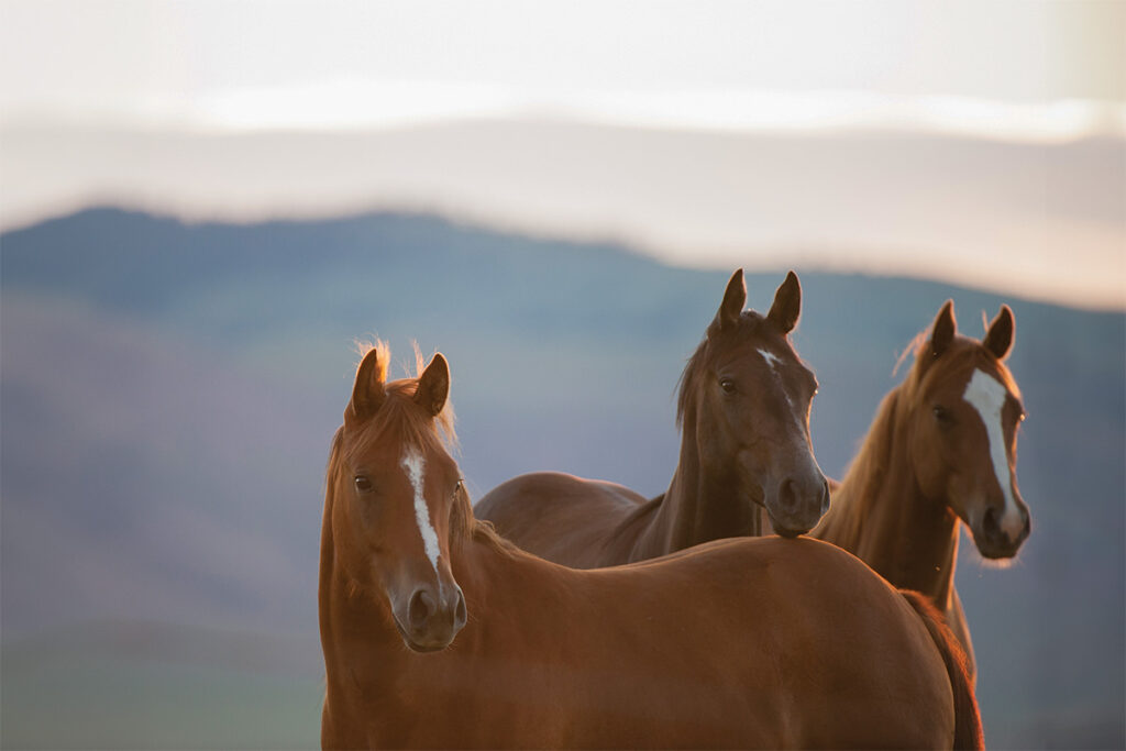
Owners often describe their horses as reactive when they spook at leaves, a gust of wind, a mouse, or something invisible to everyone else. But in some cases horses could be deemed reactive when their immune systems go haywire, resulting in disease. Fortunately, auto-immune diseases occur infrequently, which means most horses avoid their serious effects.
Scientists define autoimmune diseases as those where the immune system targets the body’s own normal proteins, causing damage and/or organ dysfunction. In contrast they refer to secondary immune-mediated diseases as the body’s excessive inflammatory reactions to foreign materials such as drugs, supplements, viruses, bacterial toxins, or vaccines.
“Key examples of autoimmune diseases include various skin conditions, such as pemphigus foliaceus and immune-mediated drug reactions that occur when the immune system builds a response to a specific drug, and the antibodies produced against the drug end up causing damage to the animal,” explains Rosanna Marsella, DVM, Dipl. ACVD, a professor at the University of Florida’s College of Veterinary Medicine, in Gainesville.
In this article we’ll talk about some of the rare autoimmune diseases based on organ system, including skin, blood vessels, and the gastrointestinal tract. But be aware that immune-mediated diseases can also affect many organ systems, including the joints (immune-mediated arthritis), muscle (immune-mediated myositis), and eyes (recurrent uveitis).
The Skin: Immune-Mediated Dermatopathies
Among rare immune-mediated skin conditions, pemphigus foliaceous ranks as one of the most common primary autoimmune disorders in horses. This condition occurs when the horse’s body produces antibodies—molecules generated by the immune system typically in response to a bacterial or viral infection—that begin attacking normal proteins located in the skin.
“Sometimes a drug or vaccine can trigger the immune system, but other times the cause is idiopathic (unknown),” says Marsella.
The proteins the immune system attacks, called desmosomes, anchor individual cells together in the skin’s epidermal layer. When the horse’s errant antibodies destroy desmosomes, it compromises the epidermal layer, resulting in the formation of superficial pustules and blisters.
Horses with pemphigus foliaceous have pustules anywhere over the body but often appear in the periocular (around the eye) and muzzle areas. But the pustules often burst and are therefore transient, quickly replaced with crusting. Owners often report a repeating cycle of pustules to crusting lesions accompanied by dullness, lethargy, and anorexia in their horses during the worst periods. Severity varies on a case-by-case basis, with some horses covered in lesions and others with lesions limited to small areas, such as the coronary band or face. Many horses become pruritic (intensely itchy) and can injure themselves through scratching and rubbing.
“Most patients are systemically ill in addition to simply having skin lesions,” says Marsella. “And the systemic illness is frequently the biggest consideration because they are lethargic and febrile. If skin lesions are extensive, then secondary skin infections are the next big concern. Quality of life is a definite issue, and most horses founder (develop laminitis where the coffin bone has rotated) with the treatment (prednisone) so the vast majority of horses with autoimmune diseases are unfortunately euthanized.”
When veterinarians diagnose skin conditions, they need to remember that pemphigus occurs only rarely and should consider other conditions first, specifically Chorioptes mites.
To muddy the waters, horses with pemphigus foliaceous can develop secondary bacterial infections, as Marsella mentioned, and veterinarians need to treat these first before performing any diagnostics. After treating or ruling out bacterial or fungal infections and ectoparasites, they can move on to the next diagnostic step: biopsies. Ideally, they’ll biopsy active pustules because the ulcerative lesions yield little information. But if pustules are no longer present (recall it is an ever-changing cycle of pustules and crusting), then veterinarians can biopsy the crusts.
“The diagnostic lesions are in the superficial epidermis and in the crusts, which is why it is so important that owners do not clean or scrub the skin before the veterinarian arrives to perform the biopsies,” explains Marsella.
Pemphigus vulgaris, a more severe and far rarer cousin of pemphigus foliaceous, causes deeper and more extensive ulcerative tissue damage in horses. When antibodies attack the layer of cells just above the basal cell layers, it results in bullae (large blisters) rather than just pustules. Horse owners and veterinarians most often see lesions in a horse’s oral cavity first and can easily confuse them with signs of more common conditions such as equine pox virus or drug eruptions and vasculitis. But in the case of pemphigus vulgaris, horses become quite ill, exhibiting fever and anorexia. As with pemphigus foliaceous, equine practitioners will use biopsies to diagnose this autoimmune condition.
The Blood Vessels: Immune-Mediated Vasculitis
Autoimmune vasculitides (conditions involving blood cell inflammation) typically develop as secondary conditions, meaning the horse’s immune system overreacts to triggers—commonly foreign materials such as drugs, supplements, viruses, bacterial toxins, or vaccines.
“When one of those triggers enters the body, the immune system creates antibodies against the foreign materials, binding to them, making antibody complexes that lodge in the very small blood vessels,” says Rana Bozorgmanesh, BSc (Hons), BVetMed (Hons), Dipl. ACVIM (LAIM), MRCVS, associate professor at the University of California, Davis, Veterinary Medical Teaching Hospital. “These blood vessels become damaged due to overwhelming inflammation, and the tissues those blood vessels supply blood and oxygen to are also injured due to lack of blood flow.”
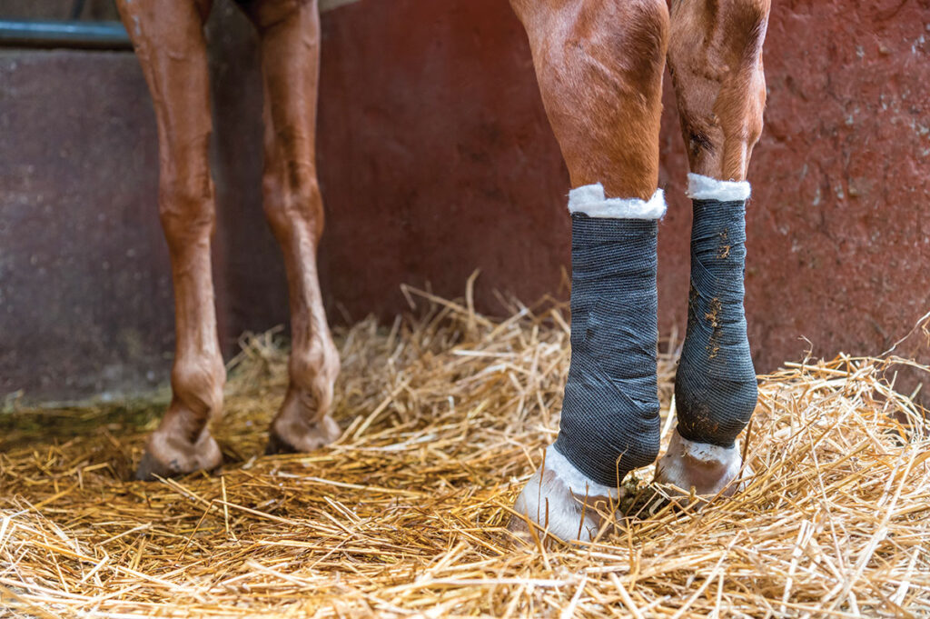
Not surprisingly, clinical signs of immune-mediated vasculitis can vary greatly depending on which blood vessels the immune complexes damaged and how severe the inflammation is. One example of an immune-mediated vasculitis is purpura hemorrhagica (PH) secondary to bacterial infections such as Streptococcus equi subspecies equi (strangles) and Rhodococcus equi or even following vaccination against S. equi. In these cases horses often have skin lesions, specifically swelling of all four limbs, exudate (seeping fluid), and even skin sloughing. These signs develop because of damaged and leaky blood vessels to the skin, causing fluid to leave the blood vessels and accumulate in the tissues.
You won’t just find lesions on the skin. Immune complexes also lodge in vessels supplying blood to the internal organs and muscles. Thus, horses might also resist movement due to muscle infarcts, show colic signs due to intestinal infarction (such as elevated heart and respiratory rates with abdominal pain), or develop internal organ damage and/or neurologic disease. In some cases horses also get nosebleeds.
Overall, these horses can appear very ill, exhibiting lethargy, depression, anorexia, and fever.
Diagnosing PH involves collecting a comprehensive history, identifying the trigger (e.g., recent infection), and ideally taking biopsies of the skin lesions, if present, and finding they show necrosis (death) of the blood vessels. A blood titer often supports the diagnosis when strangles appears to have triggered the condition.
“The most common vasculitis I see is UV-induced leukocytoclastic vasculitis in the pasterns of horses with white legs,” Marsella explains. “These horses develop ulcerative lesions, which progress to crusting and edema. It is the single most common cause of pastern dermatitis in horses with white socks that I see at the University of Florida.”
“Many drugs have been associated with the development of vasculitis in horses, including a number of antibiotics,” says Bozorgmanesh. “Diagnosis of a drug-induced vasculitis is often presumptive and based on the temporal association between the onset of signs of vasculitis and the use of the offending drug. When signs of vasculitis are identified, all drugs should be immediately discontinued while their potential association is investigated.”
The Gastrointestinal Tract: Inflammatory Bowel Disease
Inflammatory bowel disease (IBD) falls under autoimmune conditions, though experts still struggle to pinpoint how it develops in horses. However, based on the data available, they suspect an abnormal immune reaction to bacterial, viral, fungal, parasitic, or dietary antigens could lead to bowel inflammation and the clinical signs associated with IBD.
“Due to the infiltration of inflammatory cells into the wall of the intestines, horses with IBD often have impaired nutrient absorption,” says Kathryn Timko, DVM, MS, Dipl. ACVIM (large animal), assistant professor at The Ohio State University College of Veterinary Medicine, in Columbus. “In some horses this may result in chronic weight loss, colic, and ventral edema—fluid accumulation on or under the abdomen—caused by increased permeability of the intestinal mucosa as well as impaired protein absorption from the gastrointestinal tract. Horses can also present with diarrhea.”
Based on the type of cells that infiltrate the wall of specific regions of the horse’s gastrointestinal tract, IBD can be classified into subtypes. Examples include granulomatous enteritis, lymphocytic-plasmacytic enterocolitis, and eosinophilic enterocolitis, which in turn is further subdivided into various types.
Diagnosing IBD and assigning a classification starts with a suspicion based on clinical signs (described above) and blood work (often showing hypoalbuminemia, or a low protein level), along with ultrasonography revealing thickened intestinal walls. A veterinarian can only make a definitive diagnosis via full-thickness biopsies of the intestinal wall. These full-thickness intestinal biopsies, however, can only be performed when the horse undergoes exploratory surgery, which can occur in cases of severe colic.
“Most of the time we are attempting to diagnose IBD in horses … not in need of colic surgery and therefore we can only collect smaller biopsy samples from the duodenum and/or the rectal mucosa,” says Timko.
Practitioners can collect duodenal biopsies during a routine gastroscopy, but these samples are small and only reveal abnormal cells if the disease affects the duodenum. When other regions of the intestine are involved, the biopsy fails to confirm a diagnosis.
“Rectal mucosal biopsy samples are larger; however, there is no standardized histologic interpretation protocol for the evaluation of these samples, thus making a diagnosis of clinical disease challenging,” says Timko. “Similar to the duodenal biopsy, if the horse’s rectal mucosa is not involved in the disease process, the abnormal cell population will not be present, and the veterinarians can miss the diagnosis.”
As for other approaches, “there are reports of performing a standing flank laparotomy to assist with attaining a full-thickness biopsy of the gastrointestinal tract, but this procedure is not routinely performed for this purpose,” adds Timko.
Before jumping on a diagnosis of IBD for a horse with chronic weight loss, Timko says it’s important to rule out more common conditions, including dental disease. These can include poor or inadequate nutrition and caloric intake as well as parasitic disease such as cyathostomiasis (damage to the colon wall caused by encysted small strongyle larvae).
Corticosteroids Key to Managing Immune-Mediated Diseases
Whether the disease affects the skin, blood vessels, gastrointestinal tract, eyes, or joints, veterinarians typically treat it with prednisolone (because horses do not metabolize prednisone) or dexamethasone (with different formulations and durations of action that can be selected).
They must use high doses of steroids (starting at 2.2 to 4.4 milligram per day, then tapering down) to treat dermatologic conditions, often for prolonged periods of time to induce remission.
Similarly, the immune-mediated vasculitides such as PH call for several weeks of immunosuppression with corticosteroids. Early and aggressive treatment often leads to successful outcomes, with mortality rates reaching 7.5%.
“The overall prognosis for all immune-mediated vasculopathies, however, is only fair long term,” says Bozorgmanesh.
What makes these cases difficult? Managing them starts with finding the inciting cause, which proves challenging, and removing it before suppressing the inflammation. Treating with steroids in the face of an infection—while it is still active—raises concerns and puts veterinarians in a tough spot.
Veterinarians can also treat IBD with glucocorticoids—usually prednisolone or dexamethasone.
“Each case is different in how long they will require treatment, but many cases will need a few weeks and involve slowly tapering the dose,” says Timko. “Some horses may also require lifelong therapy or periods of treatment when their symptoms return. Many horses are also treated with anthelmintics (medications used to treat parasitic infections) in addition to corticosteroids.”
In some cases equine practitioners prescribe azathioprine, which they use to suppress horses’ immune systems, particularly when corticosteroids would be inappropriate; think insulin dysregulation or laminitis.
“Sometimes azathioprine has been used in addition to corticosteroids if the horse is not responding to steroids alone or if you are trying to give them a lower dose of steroids due to secondary conditions in which steroids are contraindicated,” Timko says.
Take-Home Message
Autoimmune diseases can be challenging to diagnose; veterinarians often must rule out other conditions first. Treatment typically involves steroids, which can increase the risk of laminitis in horses with insulin issues and doesn’t always lead to a full recovery. The good news: These diseases remain rare.