Limb Positioning for Assessing Joints via X Ray (AAEP 2012)
Lower limb radiographs can help practitioners uncover valuable information about bones, joints, and joint balance in equine athletes, but Colorado State University (CSU) researchers have determined the usefulness and accuracy of this information depends largely on how the horse stands during X ray capture.
“Imbalances in certain joints such as the interphalangeal joints, also called the pastern and coffin joints, can influence gait, biomechanics, and soundness in horses,” said Erin Contino, DVM, MS, of CSU’s College of Veterinary Medicine, in her presentation at the 2012 American Association of Equine Practitioners Convention, held Dec. 1-5 in Anaheim, Calif.
Imbalances can occur for several reasons, such as the horse’s natural conformation, the type of shoe he is wearing, or how he is shod. Imbalances in the foot where one heel bulb is “higher” than the other, for example, can impact the balance of the joints higher up in the limb, change how the horse bears weight, and even how he moves. In fact, notes Contino, foot imbalances can influence gaits, biomechanics, and soundness.
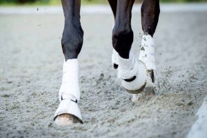
Further, Contino and colleagues suspected that how the horse’s foot is positioned while taking radiographs can influence joint balance, making accurate X ray interpretation difficult if the foot is not positioned appropriately
Create a free account with TheHorse.com to view this content.
TheHorse.com is home to thousands of free articles about horse health care. In order to access some of our exclusive free content, you must be signed into TheHorse.com.
Start your free account today!
Already have an account?
and continue reading.

Related Articles
Stay on top of the most recent Horse Health news with


