
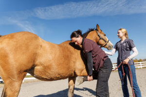
Visualizing Joints During Surgery
Arthroscopic surgery in horses is commonly performed to remove abnormal pieces of cartilage, called osteochondral (OC) fragments, from the surface of joints. Visualization through an arthroscope is typically aided by
- Topics: Article, Other Veterinary Technologies
Arthroscopic surgery in horses is commonly performed to remove abnormal pieces of cartilage, called osteochondral (OC) fragments, from the surface of joints. Visualization through an arthroscope is typically aided by distending the joint with sterile fluid. In certain joints (such as the fetlock), fluid distention is often insufficient for visualization.
“The fetlock joint is different from other joints because OC fragments tend to be obscured by the joint capsule with its synovial villi (finger-like projections of synovial tissue lining the joint),” says Nick Jansson, DVM, PhD, Dipl. ECVS, chief surgeon at Skara Equine Hospital in Skara, Sweden, and author of a study examining arthroscopy of the fetlock. Jansson’s objective was to evaluate CO2 gas distention for OC fragment removal in the fetlock joint. It was hoped that CO2 gas would help flatten the problematic villi and improve visualization.
Over two years, 26 horses were admitted for arthroscopic surgery of the fetlock. After anesthetizing each horse, the affected joint was distended with sterile fluid. An incision was made to allow passage of the arthroscope. Fluid was allowed to drain while CO2 gas was gently pumped into the joint to distend it. The surgeon could then visualize the inside of the joint through a camera attached to the scope. The fragment(s) was grasped, separated from the joint surface, and brought out of the joint with a pair of forceps. Finally, a padded bandage was applied and radiographs taken to confirm complete removal of the fragment(s). The horse was then restricted to stall rest (two weeks) and small paddock rest (four weeks) before being returned to work.
The gas distention technique proved to be an overall success, providing a clear view of the fetlock joint while flattening the synovial villi. In five horses, blood from the incision entered the distended joint and temporarily obscured the field, but “a small amount of fluid flushed through the tip of the arthroscope was sufficient to control the problem,” said Jansson. While surgical time was not shortened with the gas distension method, Jansson said, “This technique applied within the fetlock presses away the joint capsule, therefore improving visibility and facilitating fragment removal
Create a free account with TheHorse.com to view this content.
TheHorse.com is home to thousands of free articles about horse health care. In order to access some of our exclusive free content, you must be signed into TheHorse.com.
Start your free account today!
Already have an account?
and continue reading.
Related Articles


Stay on top of the most recent Horse Health news with


