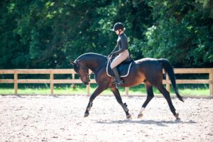
UC Davis Acquires First Equine PET Scanner
The University of California, Davis (UC Davis), William R. Pritchard Veterinary Medical Teaching Hospital recently acquired a positron emission tomography (PET) scanner, becoming the first veterinary facility in the world to utilize the imaging technology for equine patients.
In association with the UC Davis School of Veterinary Medicine’s Center for Equine Health (CEH), the hospital will launch use of the PET scanner in the summer of 2016. The unit has been acquired for research and clinical studies on lameness diagnosis in horses.
While most other imaging techniques provide “morphological” information (identifying changes in size, shape, or density of structures), PET is a “functional” imaging technique, observing activity at the molecular level and detecting changes in the tissue before the size or shape is modified. Once morphological changes have occurred, PET can tell whether the changes are still active or not.
“In practicality, that means two things,” said Mathieu Spriet, DVM, MS, Dipl. ACVR, ECVDI, a UC Davis veterinary radiologist. “One, PET can detect lesions that other advanced modalities do not identify, and two, it can tell us if a lesion—identified with another modality—is a significant injury or not
Create a free account with TheHorse.com to view this content.
TheHorse.com is home to thousands of free articles about horse health care. In order to access some of our exclusive free content, you must be signed into TheHorse.com.
Start your free account today!
Already have an account?
and continue reading.
Related Articles


Stay on top of the most recent Horse Health news with