
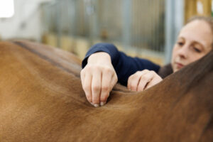
When veterinarians and hoof care professionals studying equine anatomy dissect hoof cracks in cadaver limbs, they usually slice down the middle of the fissure and look at the cross section for clues to the biomechanical and pathological causes. Longtime hoof trimmer Paige Poss thought she might be missing something in examining hoof tissue this way, so she did it differently.
“I thought, ‘This time I’m going to cut a window,’” she said. “‘I’m going to take off the front wall and look at the anatomy inside.’”
What the self-described amateur anatomist and photographer found by examining tissues this way changed her perspective on persistent hoof cracks in horses. She shared what she learned with veterinarians and farriers at the 11th annual Northeast Association of Equine Practitioners (NEAEP) symposium, held Sept. 25-28 in Saratoga Springs, New York.
Poss showed detailed images of damaged tissue, with a goal of helping hoof care professionals understand why it might be difficult to fix some cracks, and when and why you might want to consider calling the veterinarian for diagnostic imaging.
“Visually you will get an idea of why some of these cracks persist or reoccur,” she said. “Then (you’ll) gain understanding on how or even if to treat,” along with how to communicate the expectations to owners. She added the disclaimer that most of her clients are owners of older horses without major athletic demands.
“I find cracks to be one of the most interesting things,” she said. “Owners are often way more concerned over cracks that rarely cause lameness than they are over issues that are truly detrimental to the horse, such as major hoof imbalances. At the lower levels of performance, cracks are generally not a problem, and a lot of these athletes do well, though horses working at speed or with greater athletic demands might struggle with those cracks.
“Everybody wants the hoof wall to be blemish-free and beautiful,” she continued, “and being able to communicate that a lot of horses aren’t lame on those cracks and that sometimes there is a reason that it’s always going to be there is … nice (information to have) to tell owners.”
Toe Cracks


“You have changes to the laminae, changes to the corium, but when you look one step deeper, you have clear changes to the coffin bone,” she said. What isn’t clear is whether the pressure of the fused, hardened PELs causes the bone loss, or vice versa.
“These internal changes cause the outer hoof wall to not have good attachment in this area,” she added. She likens this process to a rotten or otherwise damaged wall stud that a builder covers with new drywall. The outside looks great, but everything falls apart when you add pressure.
Those internal shifts are helped by good balance and a strict trimming and shoeing schedule, she said. “Sometimes I have owners push their appointment out one week and (by then) the crack is back and as big as it was when it started,” she said.
Double Cracks


In this hoof the epidermal laminae “spikes,” as she referred to them, were significantly large and hard. “It appears they were more invasively pushing on the coffin bone and were the possible cause of the extensive bone loss,” she said.


Quarter Cracks

Next, she showed a quarter crack to the audience, then compared it to a severe quarter crack with coffin bone damage beneath it. “There’s so much tissue damage, you’d have to be very careful on how you handle (the latter crack),” she said, noting that “you’re always going to be fighting this.”
The hoof wall and the sensitive laminae beneath appeared to fold inward.
In many cases the farrier or veterinarian might use a plate or wire to stabilize the crack. But Poss cautioned that more damage to the underlying structures could occur if the crack is stabilized incorrectly.
She said she’d like to have the opportunity to diligently photograph more of these cracks because she believes “the more we see and share, the better we can prevent or treat in the future.” Some quarter cracks are relatively benign and straightforward, whereas others are tricky and cause lameness.
Heel Cracks

Poss has never had a heel crack to dissect, so she showed a photo of a heel crack in a live Thoroughbred.
She theorized that such gaps are caused by pressure on soft tissue only; there’s no bone involvement.
Solar Cracks

Bar Cracks

She moved on to showing cracks in the bars of the foot, noting that the bars are actually hoof wall but are on the bottom of the hoof on either side of the frog.
Poss showed a foot she perceived as unusual—it was very tall, unlike many of the feet she sees—but she didn’t think much of the bar crack until she dissected it.
Beneath the crack she found a 90-degree deviation of the laminae. The other bar crack she showed didn’t have as much involvement.

Take-Home Message
Poss consistently saw significant damage beneath the cracks in her dissection studies, both to laminae and to the coffin bone—damage she attributes to imbalances and lack of farrier care.
“I think it’s really super clear why we’re going to be managing a lot of these for the rest of the horse’s life, when you see how much bone loss is behind them,” she said. “You can see how it might be tricky … to keep the crack stabilized.
Poss said that it’s important for farriers and veterinarians to learn to communicate expectations, helping owners understand that if they’re really concerned about a crack, they need to get 60-degree DP (dorsoplantar) X rays to reveal what’s going on beneath it.
“If there is a void behind the crack, then more than likely you’re not going to be able to keep it gone for forever,” she said. And, finally, the onus is on the horse owner to stay on a super-tight 4- to 5-week trimming schedule to help keep those chronic hoof cracks under control.