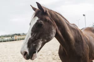
How three equine athletes returned from injury to the show ring
At some point in your horse care journey, you’ve likely ridden a beautiful round, brought your horse in from turnout, or unloaded him from the trailer and realized something was “off.” Maybe it was a lame step or the slightest bit of swelling but, either way, it prompted a call to your veterinarian. If you were lucky, the root cause was something minor that would resolve with time off and anti-inflammatories. With the more challenging cases, however, you and your veterinarian might have pursued further diagnostics to determine the cause.
After reaching a diagnosis and settling on a treatment plan, you began the arduous process of healing and rehabilitation. This stage can be trying for even the most patient equestrian. But, with a good equine care team and some time, it can be smooth and fruitful.
To see what effective rehabilitation looks like, we found three real-life examples of equine athletes that made full recoveries from their injuries. We’ll share each one’s diagnostic challenges, rehab modalities, and recovery details.
Chanel
Chanel, a 10-year-old Quarter Horse mare competing in Western pleasure, had been struggling for years with a nagging intermittent left front lameness. Her owner and veterinarian managed this with routine coffin joint corticosteroid injections for about two years. However, the injections ultimately proved to be ineffective at keeping Chanel completely sound and comfortable, so she was referred to Carrie Schlachter, VMD, Dipl. ACVSMR, who founded and designed Circle Oak Equine Sports Medicine’s rehabilitation and fitness programs and also founded Animals In Motion, a practice that focuses on integrative sports medicine, rehabilitation, and injury prevention.
“The case was pretty routine,” says Schlachter. “We nerve-blocked (used local anesthesia to numb and pinpoint the painful area) her foot, then we X rayed the area, and the X rays showed some mild abnormality in her coffin bone. We recommended an MRI so we could look at the area more deeply.”
The MRI showed that Chanel actually had two injuries to her left front foot. “The first was mild coffin bone bruising and remodeling in the area we had been looking at radiographically,” Schlachter says. “But, on the opposite side of the foot, she also had a collateral ligament injury.” Collateral ligaments are located on either side of most joints.
This was the “aha!” moment, she says: “Without the MRI I wouldn’t have known about the collateral ligament injury so, because the owners were willing to do the MRI, I was not only able to confirm my diagnosis of the bone bruising and remodeling but I was also able to see the reason for it.”
Chanel had likely been compensating for the collateral ligament injury by bearing more weight on one side of her foot, creating the bruising in the coffin bone. The injections helped initially because they suffused the area with steroids, reducing inflammation and allowing her to continue working soundly for a brief period.
With a diagnosis in place, Schlachter recommended putting Chanel in a bar shoe to support and stabilize the collateral ligament and the coffin bone. She and her team also injected the coffin joint and the collateral ligament with autologous protein solution (a biologic therapy that stimulates the body’s production of anti-inflammatory mediators and growth factors) and treated the area with extracorporeal shock wave therapy (believed to improve new blood vessel growth, recruit mesenchymal stem cells, and have pain relieving effects).
Schlachter also recommended for Chanel a controlled exercise program, which she modifies to meet the needs of different injuries and disciplines but typically involves:
- Eight weeks of stall rest with handwalking multiple times each day;
- Eight weeks of walking under saddle;
- Eight weeks of walking and trotting under saddle;
- Six to eight weeks of walk-trot-canter (or lope, as it were) under saddle with sport-specific additions (such as jumps) added at the end of this stage.
Two months post-diagnosis, Schlachter reevaluated Chanel. “At that point she was 80-90% better, so we allowed her to be walked under saddle for the next two months,” she says. “When we looked at her again at the four-month mark, she was 100% sound, so we started her on some trot work.”
Once she was sound at the canter, Chanel began working back into training. Eight months post-diagnosis she was still sound and back in the show ring. She is now free of bar shoes, and her only maintenance since recovering has been a round of hock and sacroiliac joint injections to manage normal wear and tear.
“Chanel was a wonderful patient,” Schlachter says. ”She is the picture perfect example of what a good diagnosis, good treatment, compliant owners, and a well-behaved horse can do.”
JR
Melissa King, DVM, PhD, Dipl. ACVSMR, is an associate professor at the Colorado State University (CSU) Veterinary Teaching Hospital, in Fort Collins, where she specializes in equine sports medicine and rehabilitation. King treated JR, a 16-year-old Thoroughbred who had shown as a four-star eventer. From repetitive use in his job, JR developed an insertional lesion in his deep digital flexor tendon (DDFT, which runs from the knee down the back of the leg and around the navicular bone, attaching to the coffin bone) and a second, discrete tear at the pastern level.
Diagnosing the injury was challenging due to its location. “A portion of the lesion was at the insertion of the DDF tendon onto the coffin bone,” says King. “Since we were unable to image the soft tissue structures in the foot with ultrasound, the only path to diagnosis was through MRI.”
Once King made a diagnosis, however, she performed a contrast CT scan and a bursoscopy (using a fiberoptic endoscope to guide her debriding of the lesion), followed by an intra-arterial perfusion of bone-marrow-derived stem cells (which have anti-inflammatory properties and help coordinate tissue healing). JR’s postoperative care took place at the CSU Equine Rehabilitation Center and included physical therapy, aquatic exercise, cryotherapy (cold therapy), and controlled exercise.
“Cryotherapy was initially utilized as a means of reducing inflammation to the DDFT region,” King explains. “Underwater treadmill exercise was incorporated as a means of providing buoyancy and, thus, decreasing the loading on the forelimbs while increasing joint range of motion, stride length, and muscle strength.
“JR’s rehabilitative workload increased to incorporate the use of weighted surcingles, surface changes, backing exercises, ground poles at various heights, and eccentric loading exercises aimed at increasing soft tissue extensibility in a controlled manner,” she adds.
In addition to his forelimb injuries, JR had significant back pain and muscle soreness caused by severe overriding dorsal spinous processes (kissing spines) that were evident on radiographs. The CSU team treated these areas with acupuncture, laser, mesotherapy (corticosteroid and local anesthetic injections into the middle layer of skin on the back to help relax the muscles), and daily core exercises. JR was hand-walked twice daily using a resistance band to improve hind-limb strength and stability and encourage lumbosacral (where the lumbar vertebrae meet the sacrum at the croup) flexion.
Sixteen months post-surgery, JR made a full recovery, returning to training and, eventually, eventing at the three-star level. The only maintenance he receives now is regular farrier care to maintain appropriate hoof angles and reduce stress and pressure on his hooves and legs.
“The ultimate goal of any rehabilitation program is to return an injured equine athlete to a fully functioning, pain-free state,” says King. “In order to accomplish this, therapeutic programs must be customized to each individual, addressing mechanisms to modulate pain, restore joint range of motion, enhance neuromotor control, improve muscle strength, and increase flexibility.”
Izzy
In 2019 Schlachter was called out to a nearby show to treat Izzy, a 12-year-old Warmblood mare. Izzy had been performing well in the 1.40-meter jumpers until the middle of the summer, when she walked out of her stall with a very swollen left foreleg. She’d shown earlier that day and had pulled a few rails in the class, something that was out of character for the careful jumper.
“Because the swelling, pain, and heat was obvious, we didn’t do any nerve-blocking,” Schlachter recalls. “We just ultrasounded her leg and found a significant core lesion in her left front distal check ligament (which originates at the back of the knee and attaches to the deep digital flexor tendon).”
Back at Izzy’s farm, Schlachter performed shock wave therapy and injected autologous protein solution into the ligament, then placed her in a controlled exercise program. After two months of hand-walking, Izzy’s foreleg was still puffy and painful. As a precaution, Schlachter decided to re-ultrasound her and discovered an adhesion forming between the check ligament and the superficial digital flexor tendon (which lies over the DDFT).
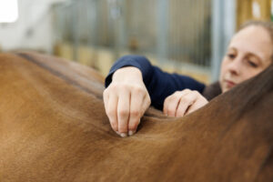
“There was a lot of swelling, which shows inflammation,” she says. “As that inflammation started to go down, it was concreting and pulling the tissues together. So, we did a second round of shock wave on the ligament, and we started implementing a routine of cross-fiber massage,” which involves using small back-and-forth strokes to help break up tissue adhesions and increase flexibility during healing.
The prescribed massage routine was concise. Schlachter instructed Izzy’s owner and trainer to pick the leg up every day, place their thumbs on the injury site, and rub across the fibers of the ligament for 60 seconds. She often recommends cross-fiber massage for check ligament injury patients.
“I see four or five cases per year of check ligament re-injury, and it’s always an adhesion in that area,” she says.
Frequent diagnostic imaging can also help ensure these are not forming at the injury site. “I like to ultrasound every 30 days during recovery when there’s risk for an adhesion, so I can keep an eye on the area,” Schlachter says. “For normal, more superficial injuries, I try to ultrasound every 45 to 60 days during recovery.”
When Izzy reached 10 minutes of trotting under saddle during her controlled exercise routine, she started getting sore again. “At that point, we looked at the rest of her body,” says Schlachter. “We found out that she was very sore in her hind end, likely from standing around. The result was that she ended up with inflammation in her hocks. We injected her hocks, dropped her back down to walking for two weeks, and then started back into the trot program, and she was fine.”
Ten months post-injury Izzy had recovered and returned to training, eventually competing in the 1.30-meter jumpers.
Take-Home Message
Being tuned into your horse’s physical and mental well-being can be key in preventing a career-ending injury. “Learn to run your hands down your horse’s legs every day so you know what’s normal and what’s not normal,” Schlachter says. “And respect your horse. If they undergo a personality change or your relationship with them is suffering, there’s usually a reason why, and it’s usually a physical one.”