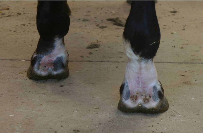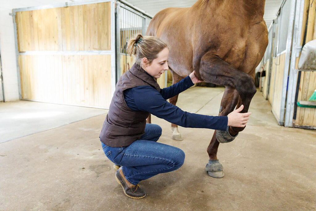
The Equine Lameness Exam Workup Explained
From palpation to nerve blocks, get an inside look at today’s lameness exam in horses in the Spring 2026 issue of The Horse.

From palpation to nerve blocks, get an inside look at today’s lameness exam in horses in the Spring 2026 issue of The Horse.

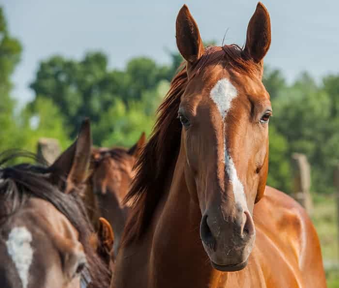
Meet Beau and follow the 8-year-old Quarter Horse’s journey from subtle lameness to return to performance.

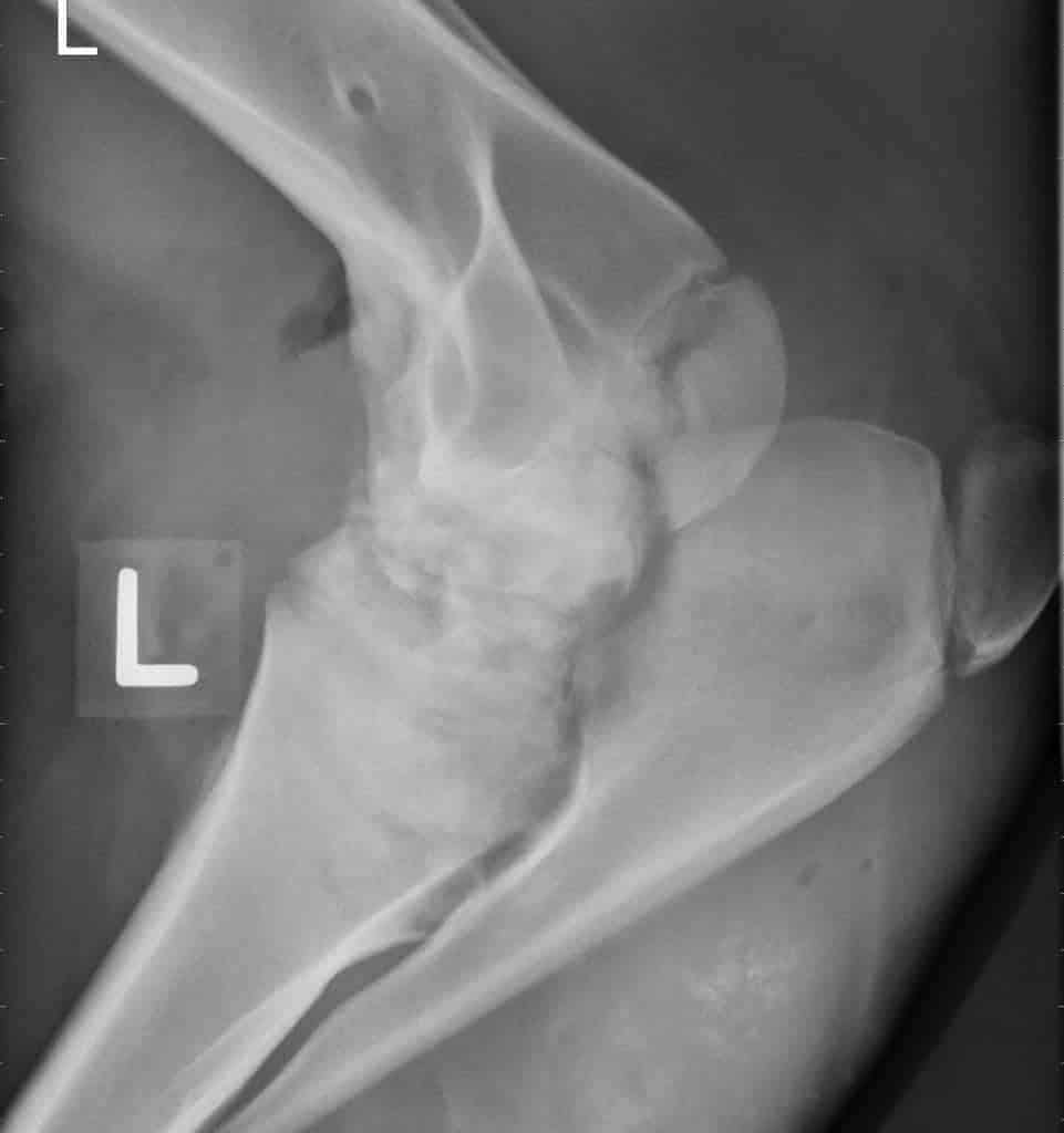
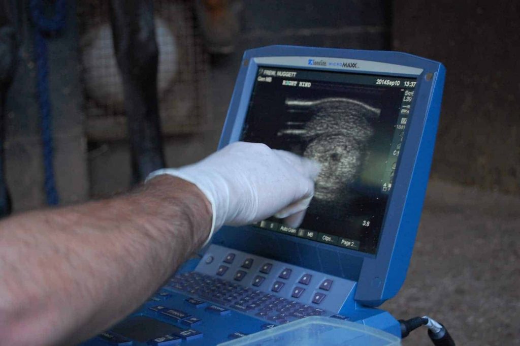
Learn what to expect when your horse undergoes advanced imaging exams using MRI, CT, and nuclear scintigraphy.

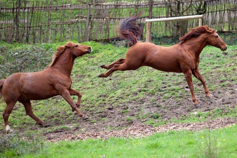
Learn how these injuries happen, how veterinarians treat them, and tips for preventing them in the first place.
Dr. Tena Ursini describes why veterinarians need to use imaging to confirm a diagnosis of equine osteoarthritis and which modalities they usually turn to first.

Diagnostic imaging results are clearer than ever, but how they will affect a horse’s performance career isn’t always evident.

Researchers described the most common abnormalities seen on imaging in nonlame show jumpers—problems that could lead to lameness in the horses’ future.

Dr. Katie Ellis and Dr. Howland Mansfield discuss what imaging modalities veterinarians might use to accurately diagnose joint disease in horses.

Two experts review how MRI, CT, and PET can provide precise answers for equine practitioners, contributing to more specific treatments and better prognoses.
The racing industry is leading the charge in identifying at-risk athletes before catastrophe occurs.

Read about the significance and prevalence of these injuries and how veterinarians diagnose them.

Getting to the root of podotrochlosis is an ongoing process. Learn about risk factors for the disease and how veterinarians diagnose it.

Veterinarians consider MRI the gold standard for diagnosing equine musculoskeletal injuries. Learn more with this visual guide.

New imaging techniques might make examining abnormalities of the fetlock easier for equine veterinarians.
Four diagnostic imaging experts share insight that can help owners and veterinarians with the diagnostic process.
Advancements in equine imaging have made it possible for veterinarians to better understand the anatomy and pathologies of their patients.