A Fresh Look at the Causes of Navicular Disease

Getting to the root of podotrochlosis, one of the most common causes of lameness, is an ongoing process
In recent years veterinarians and farriers have made big strides forward when it comes to helping horses affected by podotrochlosis (historically called navicular syndrome). Once thought to be the end of an athletic horse’s career, degeneration of the navicular apparatus is something these professionals are now well-equipped to address (see TheHorse.com/navicular). Pinpointing the exact causes of the condition, however, remains a work in progress.
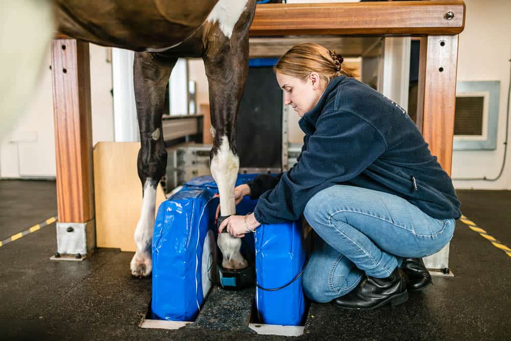
Advances in diagnostic imaging, notably magnetic resonance imaging (MRI) technology, have enabled veterinarians to gain a greater appreciation of the condition’s complexity. With increased understanding has come the realization that what we’ve labeled “navicular syndrome” for centuries is, in fact, a condition involving a number of structures beyond the navicular bone.
Affected Tissues

Podotrochlosis refers to inflammation of the podotrochlear anatomy of the horse. ‘Podo’ means foot, and ‘trochlear’ designates a pulleylike apparatus. This shift in terminology occurred as it became clear the navicular bone is not the sole culprit in these cases. Other vital podotrochlear structures often contribute to the problem. A nonexhaustive list includes the navicular bursa, coffin joint, lower part of the deep digital flexor tendon (DDFT) and its insertion on the underside of the coffin bone, and small ligaments that connect the bones inside the hoof to one another. Put together, these structures create a pulley system that helps lift the foot off the ground, and also dissipate mechanical forces upon landing. They are designed to work seamlessly together. With every step the horse takes, the DDFT glides around the navicular bone, a motion which, in a healthy horse, is cushioned and lubricated by the fluid-filled sac that is the navicular bursa.
Risk Factors
In the development of podotrochlosis, nature and nurture are both to blame. Some predispositions are inherited, others arise by way of management, conformation, and level of work. Many can, at least to some extent, be manipulated. While the exact cause of podotrochlosis is often unknown, researchers have studied and identified a multitude of risk factors over the years, says Michael Fugaro, VMD, Dipl. ACVS, owner of Mountain Pointe Equine Veterinary Services and an equine surgeon at B.W. Furlong and Associates, both in New Jersey.
“The most prevalent risk factor for podotrochlosis is genetics,” says Craig Lesser, DVM, CF, an associate veterinarian and farrier specializing in lameness and podiatry at Rood & Riddle Equine Hospital in Lexington, Kentucky. “For example, Quarter Horses, with their small feet, have the highest prevalence of the condition,” he says. Warmbloods and Thoroughbreds are also overrepresented in podotrochlosis cases. Foot conformation undoubtedly has a big impact on the amount of strain the navicular apparatus endures. Tissue damage occurs as a result of concussion when the horse is in motion.
“Although not exclusive, many horses suffering from caudal heel pain (a characteristic component of podotrochlosis) have a low heel and long toe,” Fugaro observes. This combination exacerbates the strain applied to the navicular apparatus by forcing its pulley system to work harder to lift the foot off the ground.
“In horses with such conformation, the navicular apparatus isn’t being used as designed, resulting in abnormal concussion on the bone and soft tissue structures,” Lesser explains. As with most things, improper use leads to premature wear and tear. Affected horses can display a distinctive and characteristic “shuffling” gait, especially at the trot, and might point their feet in an attempt to reduce painful weight-bearing on their heels.
Dr. Craig Lesser
Now let’s look at the “nurture” aspect of podotrochlosis development and what we can do about it. Perhaps the single factor horse owners have the greatest ability to influence is hoof care and farriery.
“Hoof care plays a large role, and there are a lot of mechanical options available to help reduce strain on the injured region to keep the horse pain-free and in work,” says Lesser. Farriers and podiatrists often initiate therapeutic shoeing when managing horses affected by podotrochlosis. But in many cases, if implemented proactively, correct hoof trimming and shoeing in the first place could preclude the need for correction. Our sources say excellent farriery shouldn’t be regarded solely as a treatment option but, rather, a way to prevent navicular apparatus degeneration. Achieving and maintaining optimal angles from the start spares the navicular apparatus undue stress.
Aside from hoof care, other factors within our control can put excessive stress on the navicular apparatus. “Unsurprisingly, the horse’s occupation can have a significant impact,” Fugaro notes— literally. Performing on hard surfaces, which further concusses the bony structures of the foot, along with making tight turns, stopping fast, moving laterally, and jumping, have all been implicated to increase the amount of impact and, there- fore, strain to the heel region, he adds.

With podotrochlosis being a degenerative condition, meaning it progressively worsens over time, experts have noted that most affected horses are between the ages of 4 and 15—prime athletic years— when first diagnosed.
Diagnostic Options
Identifying podotrochlosis in a horse is much easier today than it was just a few years ago. “When I graduated veterinary school in the late ’90s, we were limited to physical examination and basic diagnostic imaging,” Fugaro recalls. “All that could be done back then was try to localize the source of lameness with a soundness exam, diagnostic nerve blocks, and foot radiographs of horses experiencing heel pain. And the quality of radiographic images back then wasn’t what it is today.”
Another reason veterinarians were restricted in terms of imaging is due to the limitations of ultrasound in this capacity. “In the rest of the horse, ultrasonography can be used to diagnose a variety of soft tissue injuries,” Lesser says. “In the foot, however, our use of ultrasound is very limited, as it cannot image through the hoof wall, and we have limited capabilities to see true diagnostic findings through the frog and heels.”
For their part, radiographs can help veterinarians identify some degenerative changes of the navicular bone itself. But despite technological improvements over the past two decades, both radiography and ultrasonography leave veterinarians in the dark when it comes to imaging podotrochlear tendons and ligaments.
Magnetic Resonance Imaging
Fortunately, recent progress in diagnostic imaging methods has made a world of difference for podotrochlosis-affected horses, their owners, and veterinarians. Magnetic resonance imaging came on the scene of equine veterinary medicine in the late ’90s. Often the next step after inconclusive radiographic and ultrasono- graphic exams, MRI has quickly become the gold standard for diagnosing podotrochlosis. Its images give a clear view of both soft and bony structures of the foot. Fluid, a component of swelling and inflammation, also shows up.
“The advances in availability and affordability of MRI as a diagnostic tool have allowed us to better understand the complexities of the hoof,” says Lesser. “This has given us an advantage when treating foot lameness. We are now able to pick up on small changes before they become major issues that can be seen on radiographs alone.”
Research using MRI has allowed practitioners to appreciate which structures beyond the navicular bone are affected in horses exhibiting podotrochlear pain. Results from a 2007 study by Dyson and Murray showed that a significant number of horses with navicular bone disease or damage also had DDFT lesions. “There are close interactions between injuries of the navicular bone and those of the DDFT, the navicular bursa, and the coffin joint,” the authors wrote. In other words, your horse’s diagnosis might not be as straightforward as “navicular.” It could include:
- Degeneration and/or remodeling of the navicular bone, which encompasses osteoarthritis, osteolysis (bone reabsorption/destruction), sclerosis (thickening of the contours of the bone), and osteophytes (abnormal projections on the bone—i.e., bone spurs).
- Bursitis, or inflammation of a bursa, in this case the navicular bursa.
-
Tendonitis, or inflammation of a tendon, in this case the DDFT.
-
Desmitis, or inflammation of a ligament. With podotrochlosis, a number of small ligaments could be involved: the distal sesamoidean, impar ligament, or collateral sesamoidean ligaments. These hold the navicular bone in place.
- Coffin joint inflammation or arthritis.
“With access to MRI technology, we are now able to narrow down this massive ‘navicular syndrome’ term into the exact issue that is causing pain and lameness and, then, direct treatment at that issue specifically instead of using a shotgun approach,” Lesser says.
Fugaro agrees: “Having a more accurate diagnosis has allowed for better and more localized medical and surgical treatments (e.g., corticosteroid injections, shock wave therapy, bisphosphonate administration, biologics, non-steroidal anti-inflammatories, neurectomy), improved shoeing, and customized rehabilitation protocols—all of which often results in a more accurate prognosis for soundness and future athletic performance.”
PET and Bone Scans
Magnetic resonance imaging isn’t the only groundbreaking advancement for diagnosing podotrochlosis. Last year researchers (Spriet et al.) on a study published in the Equine Veterinary Journal revealed that a positron emission tomography (PET) scan showed damage to the navicular bone when all other imaging techniques—MRI included—failed to detect abnormalities. This technology offers a head start on identifying minor defects before they have a chance to snowball into major problems.
Likewise, nuclear scintigraphy (bone scan) is an imaging tool useful for catching early, hard-to-detect damage to bones. Study results suggest a significant number of horses with navicular pain but “normal” radiographs show increased scintigraphic uptake within the navicular bone during a bone scan, highlighting bone defects.
Both PET and nuclear scintigraphy technologies are much more sensitive than radiography in detecting small, early lesions of the navicular bone. Keep in mind, however, that as bone-specific diag- nostic tools, neither will be of much help if the lameness comes from soft tissues in the foot.
Final Thoughts
While a podotrochlosis diagnosis can be disappointing, says Fugaro, “it is better to have those answers and proactively treat and prevent pain that could otherwise lead to lameness down the road. Many affected horses can be safely and effectively managed.”
“There is a lot still to learn about the causes of this condition,” Lesser adds. “Prevention is key, and with time we will hopefully be able to detect early warning signs and make changes to prevent them from becoming issues in the future.”

Written by:
Lucile Vigouroux
Related Articles
Stay on top of the most recent Horse Health news with


