The trials and frustrations of navicular disease
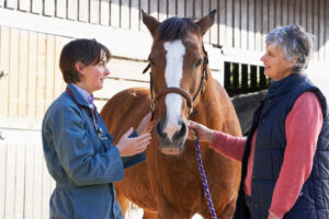
Podotrochlosis. Navicular disease. Caudal heel pain. A rose by any other name would be as … frustrating? Though it goes by several names, the condition is common among horses and frustrating for owners, farriers, and veterinarians alike. It takes a team to truly address and treat the underlying cause of a horse’s navicular-related lameness.
Navicular Apparatus Anatomy
Thirty years ago veterinarians believed any horse with navicular disease must have degeneration of the navicular bone. Decades of research and education later, we know the lameness associated with the condition might not be solely the navicular bone’s fault. Rather, many structures work in sync as the navicular apparatus. This includes the navicular bone, the navicular bursa, the coffin joint, the impar ligament, the suspensory ligaments of the navicular bone, and the deep digital flexor tendon (DDFT). One side of the navicular bone (the flexor surface) borders the DDFT. The navicular bursa “buffers” the contact between the two structures to prevent adhesions from forming between the navicular bone and the DDFT.

“The role of the soft tissue structures surrounding the navicular bone is to support the bone while the animal is standing and moving,” says Tammy Muirhead, DVM, professor of anatomy at the Atlantic Veterinary College, on Canada’s Prince Edward Island. “If one or more of these structures are affected, excessive strain can be placed on the (navicular) bone, leading to degeneration or remodeling.”
Equine practitioners classify the resulting lameness from the bone or any of the soft tissue structures as podotrochlosis, because the condition can involve any part of the podotrochlear apparatus: the navicular bone, DDFT, navicular bursa, and supporting ligaments.
Clinical Signs
The manifestations of caudal heel pain aren’t black and white. Horses with podotrochlosis display any number of clinical signs, which can make coming up with a diagnosis and treatment plan a long, challenging battle.
The lameness most often affects horses around the ages of 7 to 9, though it can appear in equids of any age. The horse often starts showing signs of lameness after a period of rest and reintroduction to work. Early in the disease, some horses “work out” of the lameness after a proper warmup. The degree of lameness might vary from day to day, which can be incredibly frustrating for owners and vets.
Horses with navicular-related lameness often display a very short-strided or “choppy” trot. They tend to stumble at a trot or even a walk and have a visible head bob correlating with one of the forelimbs. The lameness usually worsens if the horse is forced to circle or trot on a hard surface (concrete or driveways are ideal for lameness workups).
The disease usually affects both forelimbs, though one forelimb might be worse than the other. Nerve blocks (by which veterinarians desensitize the navicular apparatus) of the lame foot often reveal lameness in the seemingly “good” leg.
Diagnosis

Veterinarians can use several techniques to definitively diagnose podotrochlosis in horses. However, many “navicular disease” diagnoses are based on clinical signs alone due to lack of practitioner experience, availability of diagnostic equipment, or owner finances.
The most common imaging modality used to diagnose the condition is radiography. The veterinarian takes a series of X rays to get a detailed look at the affected areas of the navicular bone. He or she scrutinizes the images for bone surface abnormalities, the distinction between the outer cortex and inner medulla, and bony remodeling and lysis (bone loss). The vet also carefully analyzes the interface between the DDFT and the flexor surface of the navicular bone for lesions.
If you’re thinking, “I thought ultrasound was the best way to visualize tendons,” you’re right! However, due to the tendon’s close association with the navicular bursa, lesions and calcifications on the tendon can be visible on radiographs.
Some horses with caudal heel pain have few to no visible changes on radiographs. These horses might warrant a more intensive diagnostic plan. While ultrasound is a great tool for looking at tendon and ligament injuries, it can be difficult to see the tissues of the navicular apparatus due to the horse’s lower limb anatomy and ultrasound’s limited ability to penetrate the hoof wall. Researchers have found that MRI is incredibly helpful for definitively diagnosing podotrochlosis in horses. It provides the best and most complete imaging method for the equine hoof.
Trimming and Shoeing
Your farrier is the frontline against the war on podotrochlosis. Usually, corrective trimming and shoeing is the first therapy we attempt when managing horses with the condition. For this treatment to be successful, your veterinarian and farrier must work as a team to come up with the best plan for your horse.
“There are several conformation types that can experience heel pain,” says Betsy Lordan, DVM, CJF, of SRH Veterinary Services, in Ipswich, Massachusetts.
Veterinarians commonly see long-toed, low-heeled horses with these issues. “That toe acts like a lever arm and puts added stress on the navicular apparatus and the other soft tissue structures in the back half of the foot,” Lordan says, adding that shortening the toe can make a world of difference for these horses.
The orientation of the coffin bone itself can be a factor in caudal heel pain. In horses with low heels, the coffin bone can drop backward, stressing the entire navicular apparatus. This is known as a negative palmar angle (the angle the bottom of the coffin bone makes with the ground). While trims are important, horses suffering from podotrochlosis might need proper hoofwear, as well.
“There are a variety of mechanisms we can use to compensate for what cannot be achieved with the trim,” says Lordan. “A negative palmar angle can be addressed with wedges or with a rocker- style shoe, and optimal breakover can be built into the shoe itself.”
Farriers also commonly add heel wedges to help navicular horses. The elevation, in theory, relieves DDFT pressure on the navicular bone, which in itself can cause heel pain. While wedges are popular with veterinarians and farriers alike, not all horses respond well to them, causing further frustration for all involved.
Talented farriers can look at a hoof and estimate what’s going on inside, but the only way to definitively see the structures is with X rays. “Radiographs will give us the palmar angle, hoof-pastern axis (alignment of the hoof with the pastern angle), and help us determine how much hoof wall or sole we have at our disposal for the initial trim (as well as other) bony pathology (disease or damage) which may need further consideration,” says Lordan. “Radiographs give your farrier another window into the foot to use as a guide.”
Oral Medications
Non-steroidal anti-inflammatory drugs, or NSAIDs, are popular treatments for confirmed cases of podotrochlosis. Phenylbutazone (Bute) and firocoxib (Equioxx) are two potent NSAIDs commonly used to decrease inflammation caused by the condition. Usage of NSAIDs does come with risks, however, including gastrointestinal ulceration and kidney damage, so talk to your veterinarian before administering.
Firocoxib has gained popularity because it’s proven to be gentler on the horse’s gastrointestinal tract than Bute.
Bisphosphonates
Available in Europe for years, bisphosphonates are a relatively new drug in North America. After FDA approval in 2014, these drugs have gained a place in the navicular toolbox. As opposed to other medicines, bisphosphonates address podotrochlosis at a cellular level.

“The horse’s navicular bone undergoes remodeling throughout its life, not too different than our own bones,” says JD Conway III, DVM, of Conway Veterinary Services LLC, in Decatur, Texas.
Remodeling in any bony structure is driven by osteoclasts, cells that absorb bone tissue naturally. Overaggressive osteoclasts, however, can cause problems that lead to inappropriate bone lysis.
“This lytic process is thought to be driven by an imbalance of the osteoclast and osteoblast cells,” Conway explains. Osteoblasts, which are cells that lay down new bone fibers, are overpowered by the osteoclastic activity in many horses with podotrochlosis. “Bisphosphonates work on down-regulating osteoclasts by stopping their energy supply and, thus, encouraging a shift back to an equilibrium state within bone,” he says.
The two bisphosphonate products on the North American market are tiludronate disodium (Tildren) and clodronate injection (Osphos). Conway says his first experience with bisphosphonates back in 2014 was eye-opening. Within three to four weeks of administering Osphos to a horse with chronic navicular pain that was consistently mildly lame, he saw a dramatic decrease in lameness grade. Within two months, that horse had returned to barrel racing competition.
Joint Injections
The injection of corticosteroids and hyaluronate directly into a joint can offer comfort to horses of all breeds and disciplines, even old pasture pets.
Veterinarians commonly inject the coffin joint and/or navicular bursa of podotrochlosis horses. Coffin joint injections help reduce inflammation in the joint caused by disruption of the navicular apparatus. Again, the navicular bursa acts as a cushion to prevent the DDFT from abrading itself on the bone. When veterinarians begin to see radiographic changes in the navicular bone or DDFT, injecting the bursa with corticosteroids might provide significant relief.
While joint injections can provide quick relief directly to the diseased area, their duration of effect differs in every horse. Some horses’ relief can last a year or more, others six months or less. While injections provide fast and sometimes lasting benefits for lame horses, they do nothing to treat the underlying disease.
Surgical Options
Surgical options to address soundness issues associated with podotrochlosis are an ever-growing area of research. Veterinarians are publishing new techniques every few years. Here we’ll focus on the most common and researched techniques for navicular pain: palmar digital neurectomy and navicular bursoscopy.
Palmar digital (PD) neurectomy
Often considered a last resort, a PD neurectomy is a procedure to “cure” lameness in the navicular horse. The palmar digital nerves run along both sides of the limb to innervate the hoof, including the navicular apparatus. The procedure is a simple transection (cutting) of the PD nerves, essentially desensitizing the entire hoof below. When other treatments have failed to manage the lameness, a neurectomy might be the next best step. However, it does not come without risk.
Andy Kaneps, DVM, Dipl. ACVS, ACVSMR, owner of Kaneps Equine Sports Medicine and Surgery LLC, in Beverly, Massachusetts, has performed many neurectomies in his decadeslong career.
“Common risks include incomplete desensitization of the heels and painful neuroma formation,” he says. A neuroma is a disorganized bundle of nerve fibers secondary to transection that can be incredibly painful.
Furthermore, even if the procedure was successful initially, the nerve might reinnervate the area, once again causing lameness. “The positive effects may last as short as two years, but often last for many years longer, if not indefinitely,” says Kaneps. While veterinarians can repeat the surgery, the procedure might be less successful the next go-round.
Bursoscopy
A relatively novel procedure gaining traction in North America is navicular bursoscopy. It’s also considered an “end-stage” option when all other therapies have failed. The procedure (an arthroscopic evaluation) gives the surgeon a total view of the navicular apparatus. Upon visual inspection, the surgeon can debride any adhesions from the DDFT or navicular bone.
Bo Brock, DVM, owner of Brock Veterinary Clinic, in Lamesa, Texas, says patients that were chronically lame and end-stage have undergone bursoscopies with great success at his clinic. Some have even returned to work. Of course, it is not a perfect fix. “These horses always need to have shoeing manipulations or corticosteroid injections, but sometimes they’re being used again for years,” he says.
Diagnostics have improved dramatically in equine medicine, maybe even faster than treatment options. “For years, we’ve been able to diagnose (podotrochlosis) better than we can treat it,” says Brock.
But with the advent of MRI, surgeons can finally recognize and treat early signs of the condition with much more success. Brock has started performing bursoscopies on horses developing early adhesions seen on MRI. “Bursoscopy is the only thing you can do that separates the adhesion,” he says. “You have to go in physically and cut it.”
Take-Home Message
Podotrochlosis is and will continue to be a frustration for owners, farrier, and veterinarians. It can be blind to age, breed, and discipline. With the advent of advanced imaging, promising medical management, and new surgical procedures, however, our hopes for horses with the condition have never been higher.