How Anatomically Accurate Are MRIs of Horse Stifles?
Researchers found that high-field MRI provided highly detailed and anatomically accurate images of equine stifle soft tissues.

Researchers found that high-field MRI provided highly detailed and anatomically accurate images of equine stifle soft tissues.

MRI revolutionized the way veterinarians diagnose problems with the equine podotrochlear apparatus. Dr. Kyla Ortved explains its importance and when it’s worth the expense.

MRI is allowing vets to identify lameness conditions that were harder to evaluate in the past. One such ailment, most frequently found in sport horses, is osseous trauma of the long pastern bone’s sagittal groove. Here’s what they’ve learned so far about this condition.

Neck pain in horses remains challenging for veterinarians to diagnose and treat, but new options are on the horizon, one practitioner says.

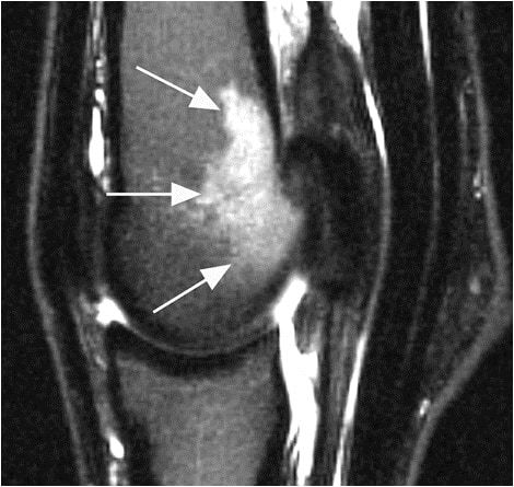
The prognosis for performance soundness in nonracing horses diagnosed with sagittal groove injury and concurrent osteoarthritis is poor, researchers found.

Ultrasound is a useful screening tool for assessing some deep digital flexor tendon lesions, but it could cause veterinarians to underestimate navicular bursa and collateral sesamoidean ligament lesions.
Lecture topics will include anesthesia, eye emergencies, sports medicine, biosecurity, foaling problems, and more.

Eight veterinarian students from Lincoln Memorial University (LMU), in Harrogate, Tennessee, spent their summer collaborating with researchers in the University of Kentucky (UK) Department of

Learn about the signs, diagnosis, and management of repetitive stress-related fetlock injuries in racehorses.
Researchers concluded that MRI can effectively show pituitary gland and pars intermedia size, as well as small details not readily visible on CT scans.

Vets can use MRI to help diagnose injuries, select treatments, monitor healing progress, and determine prognosis.
Vets use MRI to identify issues and prescribe targeted treatment to give the horse the best chance at returning to work.
Researchers found that MRI images of bone thickness could provide critical information about fracture risk.

Veterinarians discuss how they diagnose and treat injuries to the collateral ligament, DDFT, navicular bursa, and more.

Tweets and take-homes from sessions on biosecurity, equine collapse, MRI, industry issues, and more!

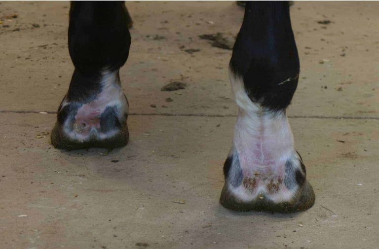
Find out how veterinarians and farriers rely on imaging to evaluate the horse’s hoof.