
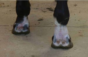
The fetlock is a complex high-motion joint that joins the bottom end of the cannon bone to the top of the long pastern (proximal phalanx); two proximal sesamoid bones sit to the junction’s rear. The long pastern’s sagittal groove (which underlies the ridge in the bottom of the cannon bone) is a common site of injury in racehorses. Researchers have documented these issues in racing Thoroughbreds and Standardbreds, and many carry a good prognosis for return to athletic function. However, how they impact other types of horses has remained unclear.
Sarah Gold, DVM, carried out the first study evaluating MRI characteristics and outcomes of sagittal groove injuries in nonracing horses, presenting the results at the 2017 American Association of Equine Practitioners Convention, held Nov. 17-21 in San Antonio, Texas. Gold is a sports medicine veterinarian at B.W. Furlong and Associates, in Oldwick, New Jersey, and Advanced Equine Imaging of Wellington, in Florida.
Gold and colleagues reviewed medical records from January 2007 through 2016 and identified 19 Warmbloods (15 geldings and four mares) with sagittal groove injuries veterinarians had examined with MRI.
Four horses had a history of chronic lameness that became acutely severe, while 15 had a history of lameness for one to nine weeks. Gold said the clinical signs the horses exhibited were variable and not specific to the fetlock. How horses “blocked” to different diagnostic anesthesia injections—designed to numb different parts of the leg to narrow lameness location—varied greatly and were not always specific to the fetlock joint. Veterinarians did not perform blocks on two horses, instead opting for nuclear scintigraphy exams (bone scan) to check for suspected fractures, with a plan for MRI based on results. All horses were lame on one leg, the degree of which ranged from Grade 1 to 3 (out of 5).
All horses had MRI evidence of a sagittal groove injury, Gold said, but the characteristics varied:
- Three had osseous cyst-like lesions (a bone defect or abnormality);
- Eight had subchondral (the layer below the joint cartilage) bone loss;
- Four had incomplete midsagittal fractures, located near the center of the bone; and
- Four had high fluid signal intensity (a lighter color on a MRI image) of the subchondral and trabecular bone of the sagittal groove (indicating fluid in the bone).
Veterinarians took radiographs in 13 horses to compare results to the MRI, she said. They found apparent abnormalities in just six on radiographs.
Furthermore, Gold said, 15 horses had concurrent fetlock joint osteoarthritis.
Practitioners treated 18 horses conservatively (i.e., non-steroidal anti-inflammatory drugs, stall rest, intra-articular autologous conditioned serum administration, etc.), and sent one horse to surgery.
At follow-up, an average of 29 months after diagnosis, Gold said six horses (31.5%) had returned to their previous use and 13 (68.5%) remained unsound, including the surgical patient. All unsound horses blocked to the fetlock joint. Two horses with incomplete fractures (“cracks” in the bone) had healed and their lameness resolved. Two horses were persistently lame and the fractures had not healed, or they developed an osseous cyst-like lesion in the sagittal groove, and three horses were persistently lame and had subchondral bone osseous cyst-like lesions.
In eight horses diagnosed initially with abnormal pastern signal, lesions were smaller, but only two of these animals were sound at follow-up. In horses with ill-defined subchondral bone loss at the sagittal groove, lesions were smaller in five horses, unchanged in two horses, and more extensive in one horse. Concurrent fetlock joint osteoarthritis was worse in three horses and remained unchanged in 12.
Researchers on previous studies saw better outcomes for sagittal groove injuries in racehorses, she said, but this could indicate these horses experience a different pathologic (disease or damage) process than sport horses. Horses in this study might have experienced chronic joint trauma and a delayed diagnosis, or might have had arthritis prior to the sagittal groove trauma, Gold said. Both could have complicated healing.
In sum, Gold said sagittal groove injuries in nonracing horses might be challenging to diagnose because of variable clinical presentations, and difficulty in predicting their outcome due to variable MRI characteristics. Further, “based on this sample, the prognosis for performance soundness in Warmblood horses diagnosed with sagittal groove injury and concurrent osteoarthritis is poor,” she said.
In addition, she emphasized that veterinarians must always evaluate the fetlock region when clinical work-up, diagnostic imaging, and treatment of the foot does not produce desired results, as fetlock injury can cause similar signs.
She noted that earlier injury detection, alternative treatments, and better rehabilitation practices might help improve outcomes. So, she encouraged practitioners to consider adding advanced imaging (such as MRI) to the examination process to allow for earlier diagnosis and treatment if no other lameness causes are identified.