Magnetic resonance imaging (MRI) offers an inside look at a horse’s internal structures. Over the past decade this modality has become the “gold standard” for diagnosing equine lameness, including those related to navicular syndrome. Standing MRI allows horses to forego general anesthesia.
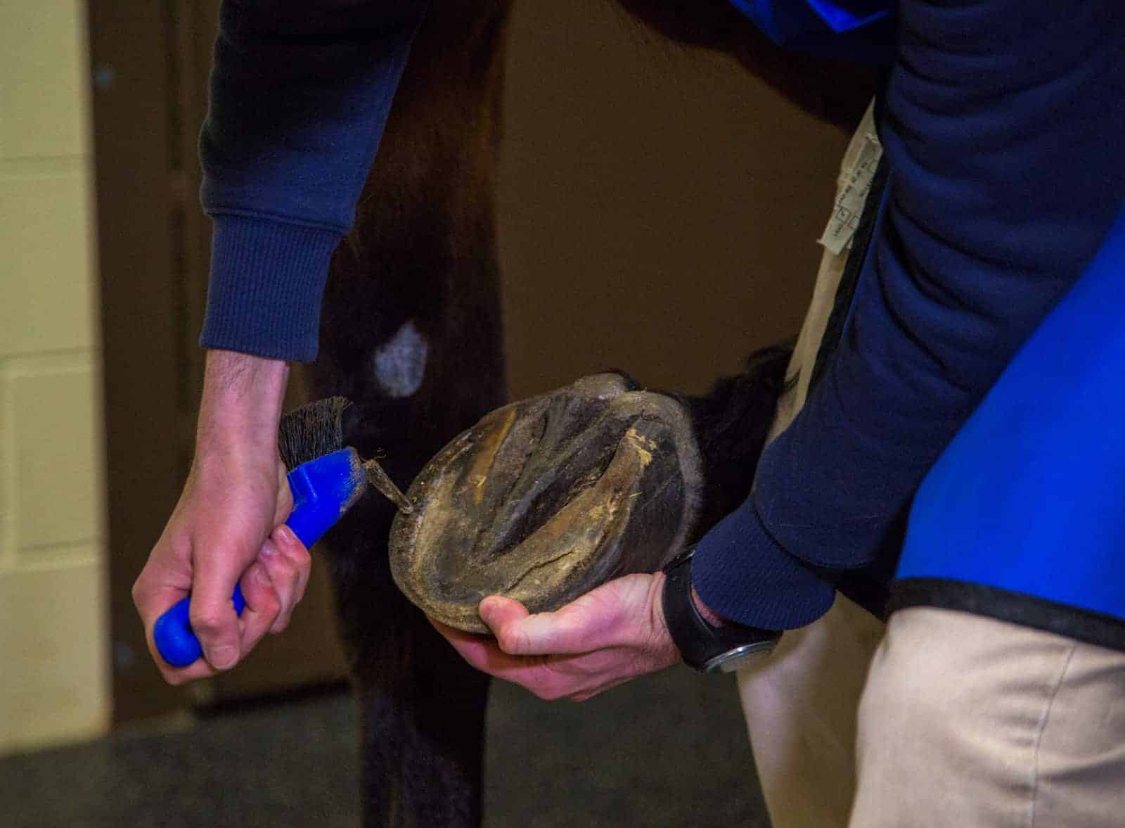
Prior to MRI: Remove Shoes
Prior to having an MRI performed, the horse must have all shoes removed and the hooves cleaned. Steel horseshoes and nails are magnetic and cannot enter the MRI scanner. | Photo: Kevin Thompson/The Horse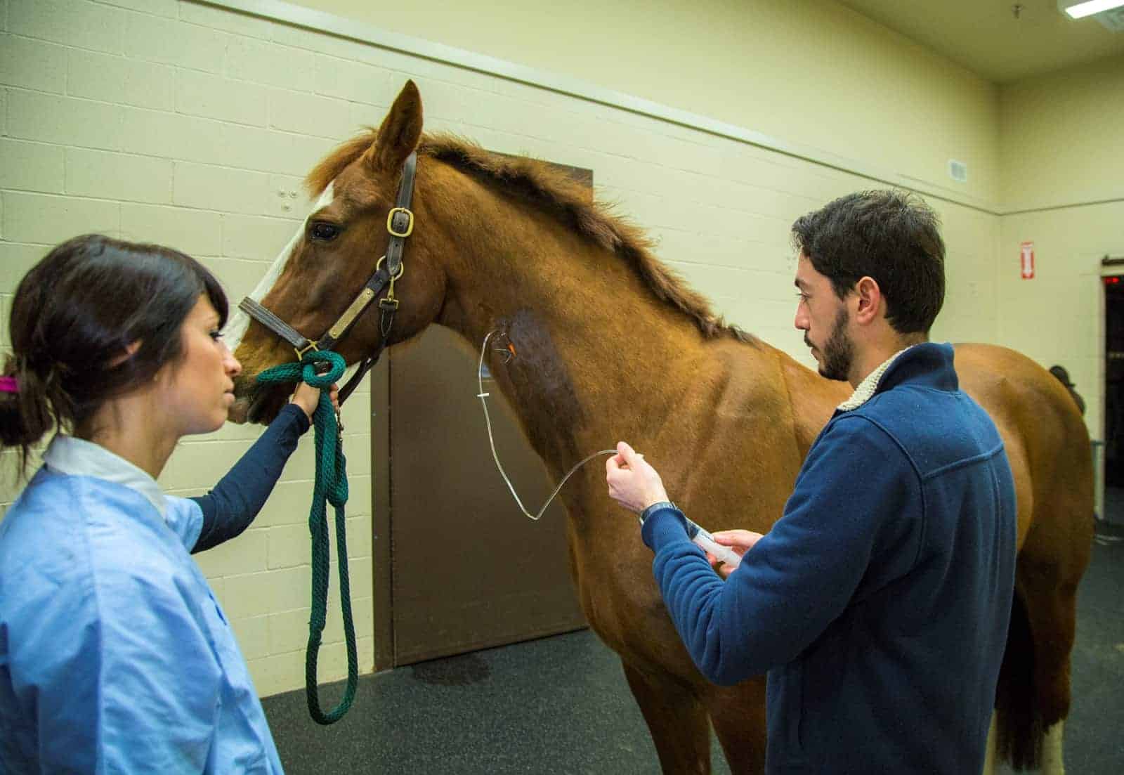
Prior to MRI: General Anesthesia
The horse is sedated prior to the MRI scan. General anesthesia, which is required for recumbent MRI, is not necessary for standing MRI. | Photo: Kevin Thompson/The Horse
MRI Room
The horse is moved from the exam area to the MRI room, which is clear of any magnetic objects. | Photo: Kevin Thompson/The Horse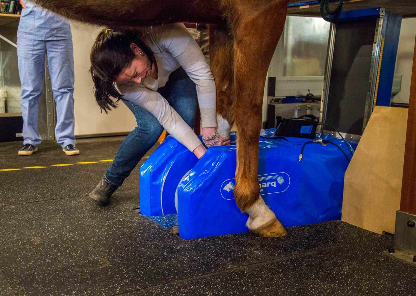
Positioning the Leg
Once the horse is in MRI room, his leg is positioned in the unit. The standing MRI scans an area about the size of a grapefruit, so veterinarians perform careful diagnostics prior to MRI to localize the area of interest. | Photo: Kevin Thompson/The Horse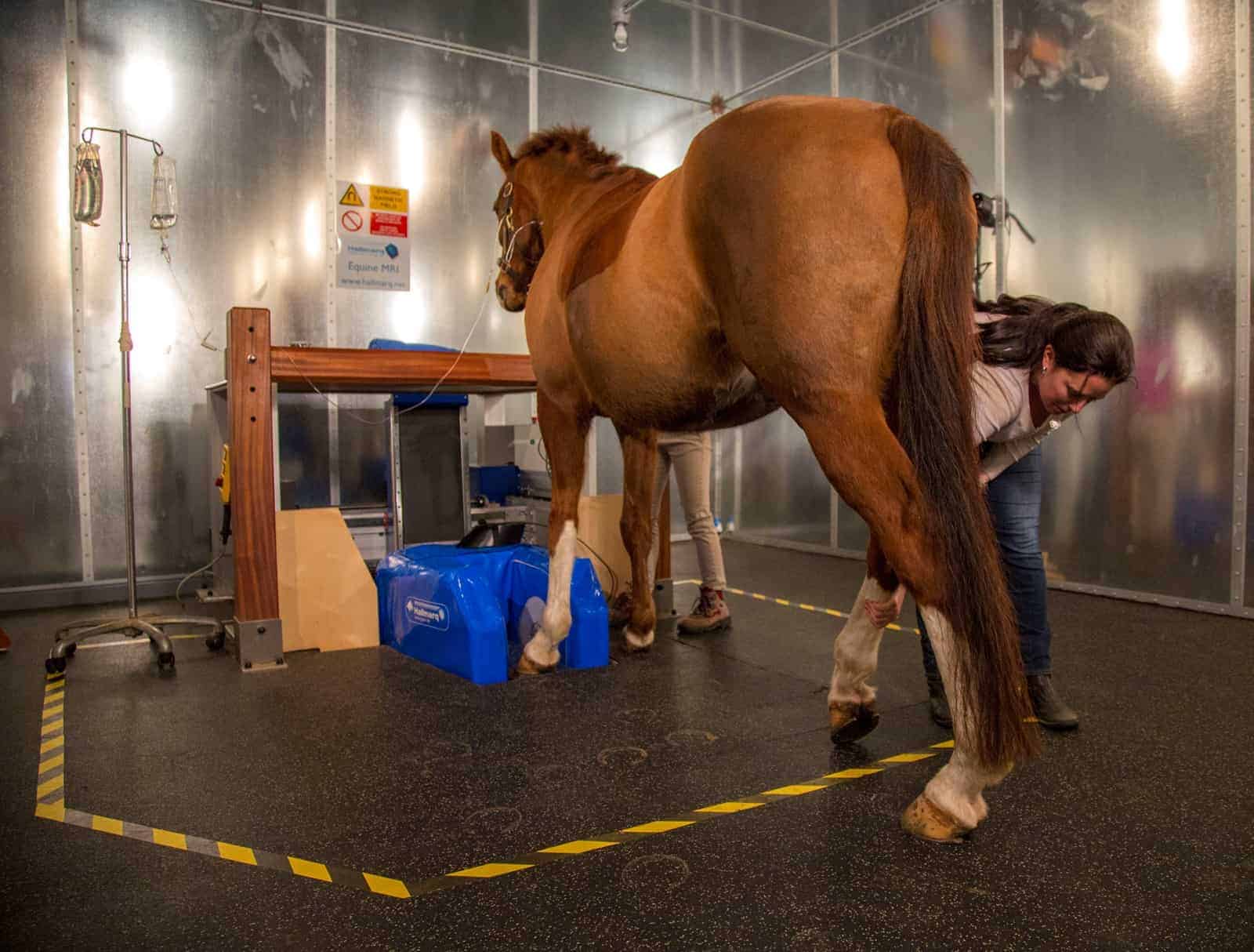
Positioning the Horse
Technicians now position the sedated horse so that he’s balanced over all four legs. This will help limit the horse’s movement during the scan. The MRI room is lined with metal, which is seen in this image, to prevent interference from external radio waves. | Photo: Kevin Thompson/The Horse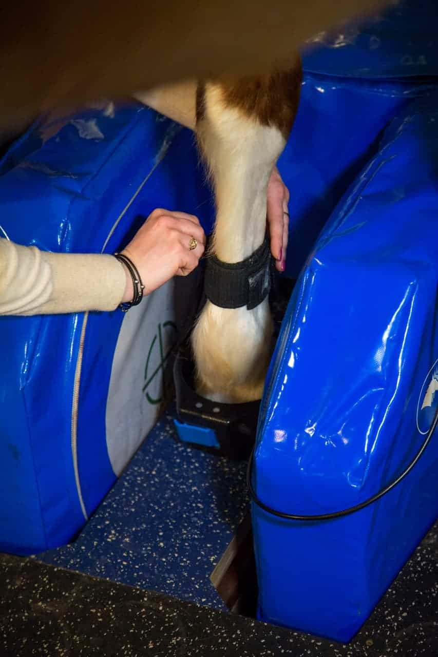
The MRI Unit
The MRI unit consists of a radio transmitter and receiver coil and a magnet. Here, a technician places the radio transmitter and receiver coil on the horse’s leg. | Photo: Kevin Thompson/The Horse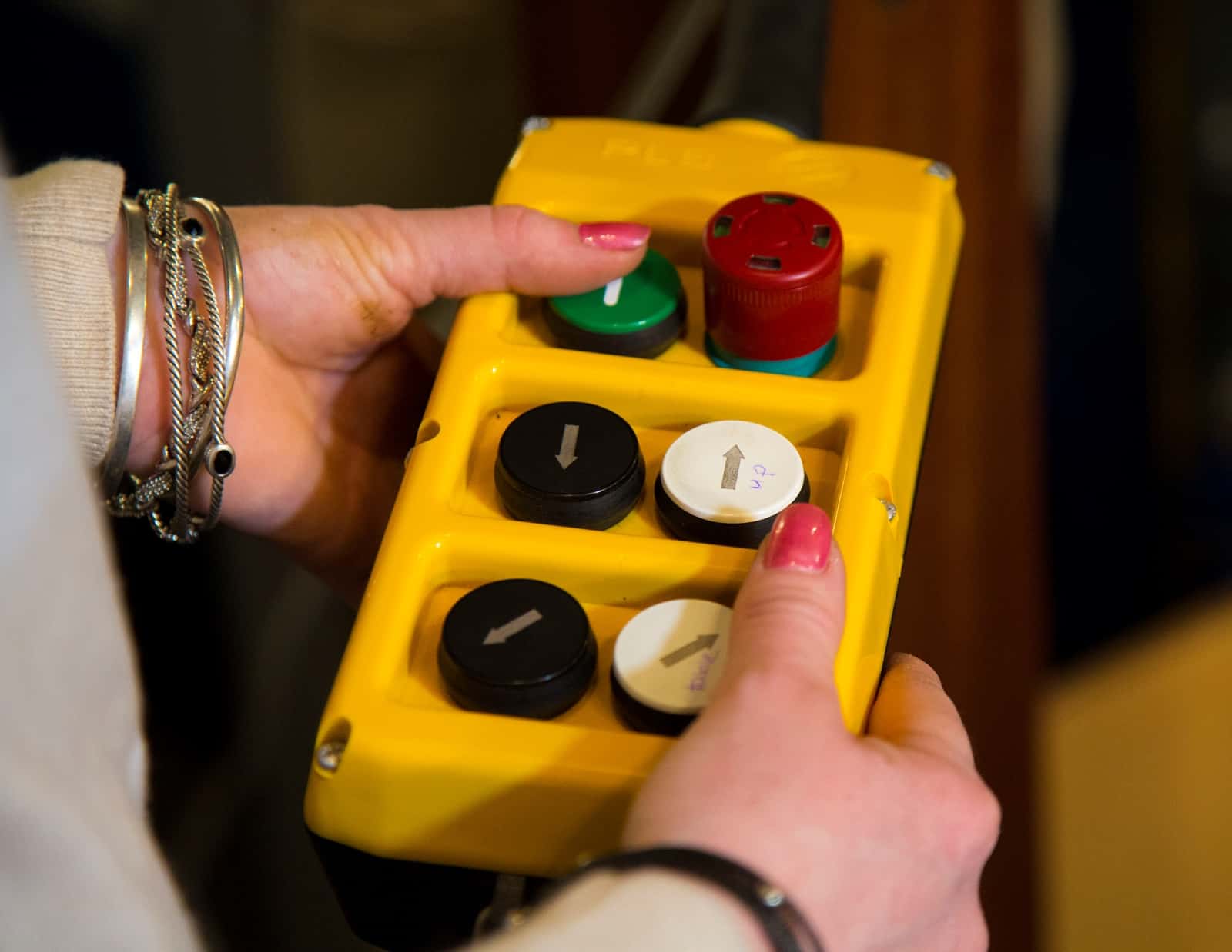
The MRI Magnet
This image shows the MRI magnet as the technician sets its position prior to the scan. | Photo: Kevin Thompson/The Horse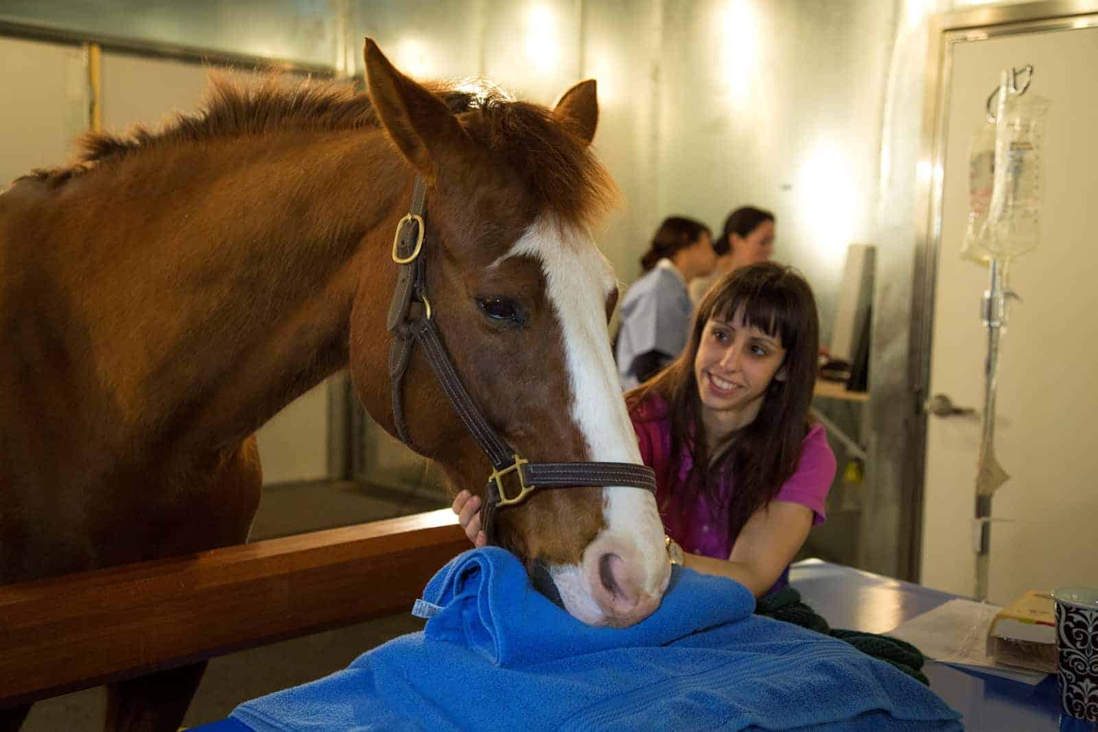
Close Monitoring
A technician stays at the horse's head during the scan, which can take 45 minutes or more. Note that the horse is wearing a halter with brass fittings, which are not magnetic. | Photo: Kevin Thompson/The Horse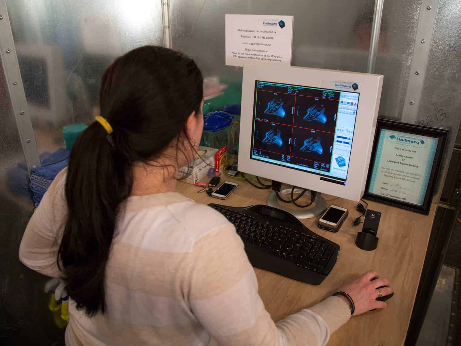
MRI Images
The MRI produces image "slices" of the foot, giving clinicians clear insight into the foot's internal structures. The images are produced digitally as soon as they're acquired. | Photo: Kevin Thompson/The Horse
MRI Images: A Closer Look
Here's a closer look of the images produced during the MRI scan. | Photo: Kevin Thompson/The Horse