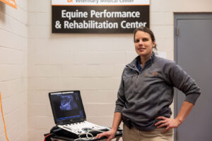
Tena Ursini, DVM, PhD, Dipl. ACVSMR, CERP
Tena Ursini, DVM, PhD, Dipl. ACVSMR, CERP, is an assistant professor in Equine Sports Medicine and Rehabilitation at the University of Tennessee, in Knoxville. Her main clinical and research interests are biomechanics and validating rehabilitation treatments, especially related to the back and topline.