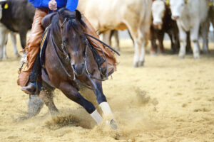
To better predict and prevent these injuries, Sarah K. Shaffer, PhD, a researcher in the JD Wheat Veterinary Orthopedics Research Laboratory at the University of California, Davis, recently investigated the tissue changes that occur prior to PSB fracture in Thoroughbred racehorses. She presented her findings at the 2021 American Association of Equine Practitioners (AAEP) Convention, held Dec. 4-8 in Nashville, Tennessee.
“The proximal sesamoid bones are essential for the integrity of the fetlock joint and, along with the suspensory apparatus, allow the horse limb to bear load during locomotion,” she explained.
Veterinarians know PSBs adapt in racehorses as a result of exercise. Because of the fetlock joint’s dense tissue, however, bony changes indicative of stress fracture have been difficult to detect and predict, Shaffer said.
What’s Going on Inside the Bone
Bone is a mineralized organic compound. Its cells can modify bone density or bone volume fraction (the amount of mineralized bone per unit volume of a sample) in response to mechanical loading and the damage repair process, explained Shaffer.
“In racehorses, the primary change that we probably see is going to be adaptation to high-intensity exercise activity that comes in the form of an increased bone volume fraction,” she said. “Functionally, this strengthens the bones and stiffens them. However, the damage repair process is really important because microdamage develops in bone tissue at physiologic loads, and the repair process first has to remove the mineralized tissue and then replace it with an unmineralized tissue called osteoid that then subsequently mineralizes. This causes a transient reduction of the bone volume fraction, which can drop the strength down a little bit.”
Shaffer explained that with stress fractures, hypothetically, a feedback loop forms where microdamage causes initial weakness before repair can occur. This temporarily reduces bone volume fraction, evident as low-density (radiolucent) areas in the subchondral bone on radiographs.
“Empirically, we know that bone strength is highly correlated to bone volume fraction,” she said.
Study Findings
Shaffer said her goal in performing the study, funded by the Grayson-Jockey Club Research Foundation and UC Davis Center for Equine Health, was to determine bone volume fraction (via micro computed tomography) and microdamage density (using a microscope) at PSB lesion sites and whether the affected areas were a stress reaction to exercise.
Shaffer’s team took medial PSB samples from 10 racehorses euthanized because of PSB fractures, as well as from 10 racehorses euthanized for different musculoskeletal injuries. Because of California’s Racing Safety Program, they also had access to each horse’s lifetime racing and high-speed exercise histories.
They found significantly lower bone volume fraction and more microdamage at locations associated with bone fracture. Bone tissue in fractured PSBs was more compacted than that from racehorses without injury. The focal drop in bone density and increase in microdamage (often called a bone lesion) is typical of a stress reaction, Shaffer said.
The compacted bone, in particular, “indicates that these horses may have been adapted to more intense training programs than the horses that were not developing the lesion in the subchondral tissue,” she said, adding that higher exercise frequency was associated with more bone loss and microcrack formation, especially in the most recent 10 months.
“This tells us that training programs likely could be modified to protect against PSB fracture,” Shaffer said. “Also, if you have the capability to monitor the lesion site, it might be a good idea to do that, because it does seem to consistently develop in horses that have a stress fracture of the PSB.”