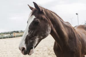
Gastric ulcers in the glandular region of horses’ stomachs appear to be associated with increased concentrations of a pathogenic bacterium—Sarcina—known to be related to slow emptying of the stomach.
Research showed that Thoroughbred racehorses had up to 450 times more Sarcina in the makeup of their glandular ulcer microbiota compared to that of surrounding healthy glandular tissue, marking the first time scientists have found such a strong connection between Sarcina and equine gastric glandular disease (EGGD). The findings suggest researchers should consider whether EGGD might be associated with poor gastric emptying, meaning food stays in the stomach longer than it should, said Sarah Voss, BVMedSci (Hons), BVM, BVS, MVM, FHEA, MRCVS, equine internal medicine clinical teaching associate in the Department of Veterinary Medicine at the University of Cambridge, in the U.K.
“Finding a pathogen that we know is associated with delayed gastric emptying in humans in horses with glandular disease is pretty interesting,” Voss told The Horse. “I suspect Sarcina probably isn’t a pathogen that’s causative of the disease. But this study definitely would support the notion that we should probably be investigating gastric emptying time in these cases.”
Researchers Sample Study Horses’ Glandular Lesions
Although scientists have made significant progress in understanding the causes, process, and treatment of equine gastric ulcer syndrome (EGUS) when it occurs in the squamous part of the stomach, they know much less about EGGD, Voss said.
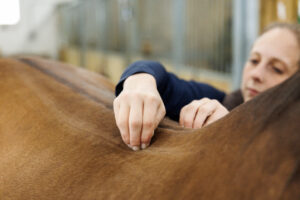
Voss and her fellow researchers performed gastroscopy on Thoroughbred racehorses to look for differences in horses with and without glandular ulcers kept in the same environment. The horses were housed at the same Scottish training yard. They evaluated five geldings and one mare, aged 2 to 7, twice and an additional horse once. All horses consumed a diet of free-choice haylage, concentrated feed, and chaff. They were not performing as well as the trainer expected them to, so she had stopped their training and requested the veterinarians scope them for possible ulcers.
After identifying ulcers in the squamous and/or glandular regions of each horse’s stomach, the scientists passed a long brush—known as a cytology brush—through the endoscope and over each glandular lesion, using a new brush for each lesion. They used additional brushes to pick up samples of healthy glandular tissue near the ulcers. This method allowed them to pick up a larger sample area than the traditional biopsy method, Voss said. It also resulted in less tissue damage while concentrating on bacteria associated with the mucosal layer itself rather than the layers of tissue beneath or the gastric lumen.
The team ran ribosomal RNA sequencing on all the glandular region samples, both healthy and ulcerated.
They found that the dominant bacteria in healthy glandular tissue were Proteobacteria, at 46.3%, about 2.5 times greater than the Proteobacteria concentration in glandular ulcers, Voss said.
The second most abundant bacteria in healthy glandular tissue were Firmicutes, at 20%, Voss said. But in glandular ulcers, Firmicutes averaged 40% of the total abundance—twice as much as in healthy tissue.
Voss said this was probably because of the steep rise in Sarcina—a Gram-negative Firmicutes bacterium. Whereas Sarcina had very minor concentrations (only 0.2%) in normal glandular tissue, it was present at up to 92.4% in glandular lesions. The rates ran particularly high in two of the three horses with EGGD, she said.
Bacteroides were the third most abundant phylum in healthy glandular tissue, representing 18.1%. In glandular ulcer lesions, however, they represented 21% of all bacteria, putting them in second place, above Proteobacteria, in relative abundance.
Sarcina Might Be Associated With EGGD
Horses might pick up Sarcina from the environment, Voss said. The bacteria are also present—albeit in much lower concentrations—in healthy mucosal samples, so they’re probably part of the normal gastric microbiota.
Even so, the large spike in concentration in glandular ulcers suggests the bacteria might somehow be associated with EGGD, Voss said. And although the study was particularly small, she added, it still hints that, given the strong link between Sarcina and poor gastric emptying in humans, horses with glandular ulcers might have stomach emptying issues.
“I don’t think this should be changing the way that clinicians are going about their day-to-day work and the way they treat these animals,” Voss said. “But I think what this should do is provide a little bit more evidence for the fact that we should be looking at gastric emptying time, because that is something that we can affect clinically.”
The samples from the horses’ stomachs showed no traces of Helicobacter, one of the main types of bacteria associated with peptic ulcer disease in humans, she said. That confirms the 2010 findings by a Danish team. Voss and her colleagues found no traces of Escherichia in the ulcers, either.
The team did not sample the squamous ulcers in this study, Voss said. All but one horse in the study had at least Grade 2 squamous ulcer disease.
“Each thing is a tiny, tiny piece of the puzzle,” Voss said. “But I think gastric emptying time might be the next tiny piece of the bigger picture.”
A study comparing the healthy and diseased equine glandular gastric microbiota sampled with sheathed transendoscopic cytology brushes appeared in the Journal of Equine Veterinary Science in April 2022.