Despite their similar names and presentations as firm swellings in the pastern region, sidebone and ringbone describe two different and unrelated processes occurring within the horse’s digit. Here’s what you should know if your veterinarian diagnoses your horse with one of these conditions.
Sidebone
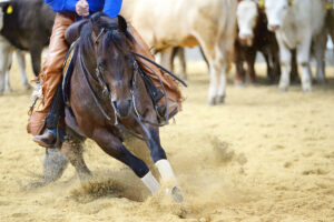
Below the fetlock are three phalanges—top to bottom, the first phalanx (long pastern), second (short pastern), and third, or distal, phalanx, also known as the pedal or coffin bone. The coffin bone is crescent-shaped, with a main body and two “wings”—or palmar processes—projecting off the back. It’s where sidebone can arise.
What you can’t also see radiographically (on X rays) are the large, thin cartilaginous wings projecting from each palmar process of the third phalanx. These ungual (or collateral) cartilages extend from the bony surface of the third phalanx within the hoof capsule to project above the coronary band, where you can feel them just above and adjacent to the heels. Their function is unclear, but they might help stabilize other structures of the digit, such as the navicular bone, to which they have several strong ligamentous attachments.
In some horses the cartilage comprising these structures gets gradually replaced with hard mineral in a process called ossification. We don’t fully understand why this happens, although theories include genetics, conformation, and concussive forces on the foot. We colloquially refer to these ossified ungual cartilages as sidebones.
The clinical significance of ossified ungual cartilages is also unclear. They’re typically regarded as incidental findings in most horses. There are, however, two main instances in which they might become a problem. First, because the ungual cartilages are mildly curved, when severe ossification occurs, the top of the now rigid cartilages can impinge upon the soft tissues sitting midline on the back of the digit, resulting in pain and lameness. To determine if sidebone is contributing to your horse’s lameness, your veterinarian can perform nerve blocks to desensitize and assess the region.
The second potential problem associated with sidebone is that once ossified, the cartilages are susceptible to fracture. This can be a significant source of lameness, particularly because such fractures commonly involve associated injury to the adjacent ligamentous structures. Some of these fractures can be subtle and difficult to detect using radiographs. Veterinarians sometimes recommend MRI for diagnosis, which also provides more information about injury to surrounding soft tissues.
If sidebone is causing a horse’s lameness, your veterinarian and farrier should work together to devise a proper shoeing and management plan.

Ringbone
Ringbone refers to osteoarthritis within the pastern (high ringbone) or coffin (low ringbone) joints. These are the joints between the long pastern, short pastern, and coffin bones. While osteoarthritis of the coffin and pastern joints is common, pastern joint osteoarthritis typically results in more extensive bony proliferation at the joint margins that might feel like regions of firm pastern swelling classically referred to as ringbone. Veterinarians usually diagnose these changes via radiographs.
Ringbone can be a source of pain and lameness, particularly if the changes are severe. Mild osteoarthritic changes in these joints are common and often managed successfully via proper shoeing and intra-articular therapeutics, such as corticosteroids, hyaluronic acid, and/or biologic therapies, while maintaining the horse in full work. If changes become severe, however, you might need to adjust the horse’s workload, particularly if medical management no longer keeps him comfortable. In these instances the severe cartilage loss within the joint (typically the pastern) might result in natural fusion (ankylosis).
Once fusion occurs, the horse might have relief from the concussive forces within the joint and improved comfort and soundness. For this reason some vets might choose to hasten the joint fusion process by injecting the joint with a substance in a procedure known as facilitated ankylosis or by surgical placement of metallic implants, known as arthrodesis. Ankylosis or arthrodesis of the pastern joint can enable the horse to return to some level of work. The same is not true of the high-motion coffin joint, where arthrodesis typically only returns a horse to pasture soundness. Your vet can recommend the most appropriate course of action for the changes occurring in your horse’s limb to provide the best possible outcome.