Sacroiliac (SI) disease disease in performance horses has many clinical signs, diagnostic methods, and causes, making diagnosis challenging for veterinarians.
Fortunately, one veterinarian recently devised a “checklist” of six clinical indicators of SI disease. He described his research and how to identify these signs at the 2014 American Association of Equine Practitioners Convention, held Dec. 6-10 in Salt Lake City, Utah.
“In general, complaints about sacroiliac disease in horses are diverse,” said presenter Rob van Wessum, DVM, MS, Cert Pract KNMvD (Equine), of Equine All-Sports Medicine Center, in Mason, Michigan. “Coming to a diagnosis can be a daunting task and often involves several diagnostic tools to exclude other sources of reduced performance.”
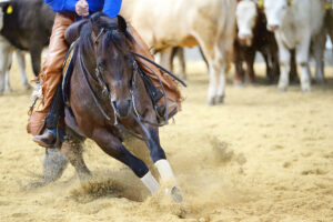
All affected horses have one thing in common, however: compromised movement of the SI region, where the spine meets the pelvis. Over the past decade, van Wessum has identified specific signs of this gait abnormality that veterinarians can test for during lameness exams.
Since 2005 van Wessum has evaluated 2,467 lameness cases with and without SI disease. During this time he noted whether each horse showed any of 26 specific disease indicators that he looked for. He then performed statistical analysis on these data to see which indicators had a significant correlation with a specific diagnosis.
Van Wessum diagnosed 327 of those horses with SI disease based on clinical exam, imaging (ultrasound and scintigraphy), and improvement post-treatment. He determined that six of his observations were significant enough to indicate SI disease. Of the 327 diagnosed cases, he said 322 (98%) had a positive score for at least three of those indicators. The indicators include:
- Tracking narrow behind “One of the first alterations of gait due to sacroiliac disease, tracking narrow behind, is often visible in walk and even more obvious in trot,” van Wessum explained. He said horses often look like they are “walking on a cord,” placing their hind feet on the same line in front of each other to help stabilize the pelvic region.
- Lateral walk Upon walking an affected horse in a serpentine pattern, the front and hind limbs on the same side move forward at the same time, similar to a pacing gait. Van Wessum theorized this is due to increased tension and decreased spinal motility.
- Haunches in/out “With sacral dysfunction, one hip is often kept slightly lower, resulting in a slight bending to one side,” he said. “This is easily observed when the horse is longed on a circle.”In this case, the observer will notice that the hind limbs don’t follow the same circle as the front limbs, with the haunches making a smaller circle than the rest of the body.
- Asymmetric tail position Horses normally hold their tails in a central position. When an affected horse walks in a serpentine, however, he will lock his tail to one side due to the SI ligament’s involvement.”When the tail is held to one side and stays to one side in the serpentine, there is a clear indication of sacroiliac dysfunction,” van Wessum said.
- “Bunny hop” canter When affected horses canter, van Wessum said they lose their normal three-beat pattern and, instead, the hind feet land together in a “bunny hop” motion to avoid the rotational forces on the pelvis. He suggested evaluating horses on both leads on the longe or while ridden and to observe the horse cantering for several minutes to rule out any hopping simply due to freshness or excitement.
- Reduced flexibility of the lumbrosacral region For this last observation, the veterinarian manipulates the SI region manually to gauge its flexibility. He or she does this by placing one hand on the point of the hip and pulling the tail toward one side, then repeating this on the other side. The veterinarian should also make the horse “tuck under” by scratching each hamstring with a pointed object.”Lateral and ventral flexibility should be symmetrical,” van Wessum said. “Clear reduction of lateral and/or ventral flexibility is a good indicator for sacroiliac dysfunction.”
Overall, van Wessum said these six tests are useful indications of SI disease and require nearly no extra effort to perform during a lameness exam.
“Statistical analysis showed that when horses do not show at least three of the indicators, it is very unlikely they have sacroiliac disease,” he said, adding that most signs disappear over time with appropriate treatment.
“Overall, this study showed how important a good clinical exam is in which the veterinarian takes time to evaluate many signs,” van Wessum concluded.