New Treatment Tactics for SDFT Injuries in Horses

Injuries to the superficial digital flexor tendon (SDFT) often heal with scar tissue that increases the chances of re-injury. But scientists are making progress in finding therapies and monitoring techniques that reduce those chances, noted an international expert at a recent veterinary meeting.
Roger Smith, MA, VetMB, PhD, DEO, Dipl. ECVSMR, ECVS, FRCVS, professor of Equine Orthopaedics at the U.K.’s Royal Veterinary College Hawkshead Campus, in Hatfield, described the challenges and available solutions related to SDFT healing during the 2022 British Equine Veterinary Association (BEVA) Congress, held Sept. 7-10, in Liverpool.
The Problem With the SDFT
The SDFT is a useful design for a swift prey species, as it provides an elastic energy source for efficient locomotion, said Smith. The problem is this efficiency is a characteristic of uninjured tendon.
“It’s a particular evolutionary development for the horse to be able to store energy and make it an efficient runner,” he said. “But as a consequence, the tendon is operating at high loads with only a narrow tolerance margin.”
Repetitive damage over time likely precedes the sudden onset of a clinical SDFT injury, he said. Once injured, the tendon becomes inflamed, which represents the start of the healing process by breaking down damaged tissue. Then this tissue is replaced through a process known as fibrosis, which produces a less elastic, scarred tendon instead of healthy, elastic tendon tissue.
“The trouble is that fibrosed tendon is dysfunctional, which predisposes the injured tendon to a very high risk of reinjury, let alone possible influences on performance,” he said.
Once the fibrosis—or proliferation—phase is complete, healing enters a final, long-lasting phase known as remodeling. While remodeling “can contribute to greater functionality,” the tendon “never gets back to normal,” said Smith.
Treatment Challenges
SDFT injuries are like Achilles tendon injuries in humans—although prolonged pain and lameness do not persist with this specific injury, he said. However, the pathology and dysfunctional healing are very similar, so new treatments might work better in both species.
Because no two SDFT injuries are identical, with injuries differing in severity and healing phase, it’s impossible to aim for a single cure-all approach. “One treatment is unlikely to be successful for all injuries in all individuals at all stages of the disease process,” Smith said.
That individual variation also makes it difficult to gather and generate evidence for different therapies, he said. Reliable studies require large numbers of study horses—around 170 at a minimum to provide a scientifically meaningful comparison between two contrasting treatment groups, which is challenging to obtain from individual clinics. Combining data from multiple practices using clearly defined outcome parameters is the way forward to accurately determine “successful healing.”
“Most studies are inadequately powered, and that’s unfortunately the reality of the situation,” he said.
Assessing re-injury rate is one of the most reliable ways to gauge successful SDFT healing to date, Smith said. But even then, the details must be clear regarding to the time frame and evaluation criteria.
“So you can see this is a highly complex picture to be able to give you some concrete evidence that one treatment is any better than the other,” he said.
Treating Phase 1: Inflammation
Because inflammation removes damaged tissue, veterinarians should not aim to stop the inflammation completely, said Smith. However, it’s a good idea to minimize inflammation to avoid damaging the surrounding, healthy tissue.
A “cheap and easy” method for this is cooling the tendon, combined with compressive bandaging, he said. Casting material placed over the top of a bandage can be a highly effective first-aid technique.
Horses should be on stall rest, of course, but more active immobilization of the leg joints can decrease the extent of damage. Certain fetlock support boots, for example, can reduce excessive loading of the tendon.
As far as medications are concerned, short-acting steroids can effectively inhibit inflammation—to the point of interfering with healing. “So they should only be used in the very acute stages, but they still have a place for those very inflamed tendons in the early stages.” Non-steroidal anti-inflammatories are less effective on SDFT inflammation, although they are useful for pain relief, he added.
Treating Phases 2 and 3: Proliferation and Remodeling
The second and third phases of SDFT healing should both benefit from physical rehabilitation therapy, which “is very crucial to educating the new tissue that’s forming a tendon,” he said.
Based on SDFT loading data from the Japan Racing Association, Smith described a “rough outline” for rehab that begins with two to four months of gradually increasing bouts of walking, which loads limbs at 60% of body weight. If the horse is comfortable after that and has good fetlock support, he can move to trotting, with loading at up to 100% body weight. After a further six months, the horse can finally begin the canter, which involves the greatest amount of loading at up to 140% body weight. At that point, the horse might also be allowed turnout time, and over the next three months this can be gradually increased back to normal work.
“The idea is that we use controlled loading of the tendon to induce better quality repair and ultimately, in the longer term, promote that remodeling process to create a more functional tendon that will be more resistant to re-injury,” he said.
New SDFT Therapies
Biological treatments such as mesenchymal stem cells (MSCs), platelet-rich plasma (PRP), and interleukin-1 receptor antagonist protein (IRAP) aim to further improve healing quality to make the developing tissue more like tendon tissue than scar tissue, according to Smith. After 20 years of use, MSCs still show the best results in SDFT repair, though they do not prevent re-injury in every case. They appear to work better in the worst-affected horses. Re-injury rates are about 50% lower in racehorses and sport horses treated with MSCs than in control groups treated with saline. Allogeneic MSCs (which come from another horse) are more convenient and less expensive than autologous (from the patient) ones, he said, and initial study results suggest they might be just as effective.
PRP treatments are showing promise in experimental conditions, “but really very, very limited clinical evidence,” he said. There’s also limited evidence about IRAP’s efficacy.
High-level laser therapy (HILT) appears to be safe, he added, “but I can’t say conclusively that it’s effective in clinical cases.” Even so, laser therapy seems to lead to a reduced lesion size, increased vascularity, and improved fiber patterns.
The Critical Need for Good Monitoring
“One of the keys to rehabilitation, where things are developing, actually, is not so much in the treatment, but in the way that we monitor these injuries going forward,” Smith explained.
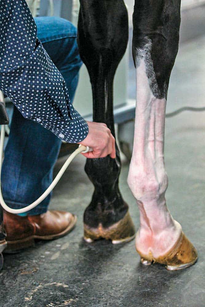
An ultrasound exam every three months, or before and after a change in exercise level, can reveal important information about how healing is progressing, based on the lesion’s echogenicity and the tendon fibers’ alignment, he said. Additional ultrasound techniques, such as the use of Doppler ultrasound (which shows the tendon’s vascularity) should gradually decrease during the healing process. “I think this is a really, really useful tool for monitoring because it’s a surrogate marker of inflammation,” said Smith.
Veterinarians can also monitor progress through high-tech gait analysis equipment or goniometers that show changes in fetlock support (the “stiffness” of the limb), which is one of the key functions of the SDFT, he said.
A new generation of “wearable tech” might provide readouts about limb function, but experts are still unsure what those readings mean in terms of monitoring tendon repair, said Smith. That’s also the case for other new monitoring technologies, such as ultrasound tissue characterization, elastography, magnetic resonance imaging (MRI), and positron emission tomography (PET). “The trouble is, we still need to understand how these outputs really reflect what is happening in the tendon and, ultimately, how we modify treatment accordingly,” he said.
Biological markers like specific genes and molecules could be the new way forward in monitoring SDFT healing, he added. Even so, results can be difficult to interpret in individual horses because markers in injured horses sometimes overlap with those found in healthy horses.
Take-Home Message
SDFT injuries can be challenging to treat, and the highest level of evidence of therapeutic efficacy is hard to come by, said Smith. New therapies are showing promise, but, for the moment, it’s the existing best practices, combined with careful individual monitoring, that provide the strongest scientific support for reducing the risk of re-injury.

Written by:
Christa Lesté-Lasserre, MA
Related Articles
Stay on top of the most recent Horse Health news with


