Researchers in Europe have reported that certain types of hock sprains in horses heal well after injury following a simple rest period, with most athletes returning to their previous activity levels after recovery.
The critical key to success in conservatively managing these tarsal collateral ligaments is good diagnostics and skilled monitoring via ultrasound, said Claudia Fraschetto, DVM, and European College of Veterinary Sport Medicine and Rehabilitation (ECVSMR) resident at the Center of Imaging and Research on Equine Locomotor Injuries (CIRALE), in Goustranville, France.
Fraschetto showed that when lesions were properly identified and followed on ultrasound, four out of every five horses treated with stall rest returned to work within six months of injury. Most of those horses reached performance levels as high as—and sometimes even higher than—those before the injury.
“The first message we want to give is that early diagnosis of these lesions via ultrasound is indispensable, even when X–rays look normal,” Fraschetto said. “And the second is that ultrasound is also very helpful in making the decision to allow the horse to return to work.”
Tarsal Collateral Ligaments: Anatomy and Injury
The hock is a very complex unit composed of several joints and bones, as well as multiple soft tissue structures, said Fraschetto. Among those structures are the medial and lateral tarsal collateral ligaments. Each ligament has two parts: a long, superficial component, and a short, deep component. The ligaments provide stability to the hock during stance and movement. Tension on the long components occurs when the hock is extended and limb weight-bearing, whereas hock flexion when lifting the limb creates tension on the short components.
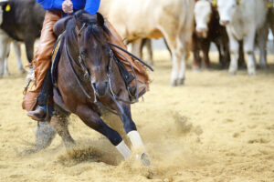
When horses sprain their hocks, it’s usually during a fall or some other kind of trauma, although sprains occasionally occur through repetitive strain, she said. Classic signs include hind-limb lameness, swelling, and pain in the affected area.
Gathering Tarsal Collateral Ligament Injury and Healing Data
Jean-Marie Denoix, DVM, PhD, founder of CIRALE, which is a unit of the National Veterinary College of Alfort (EnvA), began noticing that many of the horses treated conservatively after collateral ligament injuries of the hock could return to work, Fraschetto said.
Wanting to confirm those observations in a controlled study, she, Denoix, and their fellow researchers studied 78 horses presented as referrals at CIRALE and at the University of Liège’s Diagnostic Imaging Unit, in Belgium. The 36 geldings, 27 mares, and 15 stallions, primarily Warmbloods and Standardbreds presented with tarsal collateral ligament injuries from 2000 to 2020. About 60% of the horses had a history of trauma, such as accidents in the field, arena, stall, or trailer, or had been involved in a collision with a vehicle. At least half the Standardbreds had sustained no traumatic injuries but developed lameness that was more progressive in onset.
All the horses in the study underwent ultrasonographic and radiographic exams of the hock. Most had only one of the ligaments affected, but 21 had at least two, bringing the total number of affected tarsal collateral ligaments to 108, with a total of 111 lesions.
The short lateral collateral ligament was the most commonly affected structure, representing 44 of the 108 injured ligaments.
Ultrasound showed an increased amount of synovial fluid, known as synovial effusion, was commonly associated with these lesions and was present in 77% of the horses, accompanied by thickening or proliferation of the synovial membrane in 68% of those cases, Fraschetto said.
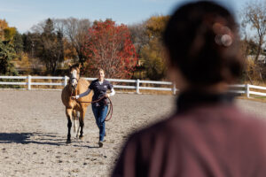
Most horses—62 of the 78—underwent conservative treatment, which involved mainly stall rest, sometimes with brief turnout in a small paddock or hand-walking, ranging from 60 to 180 days. Many of these horses also received oral or intra-articular anti-inflammatories, and some also were treated using ice therapy or corrective shoeing.
80+% of Stall-Rested Horses Ready to Work Within Six Months
The researchers found that 81% of the stall-rested horses had healed well enough to return to work within six months, Fraschetto said. Of the 44 horses for whom researchers had additional follow-up information, 86% were performing as well as or better than they had a year before their injury.
“Something that’s worrisome for both vets and owners is that persistent presence of synovial effusion, which often ends up motivating them to perform arthroscopic surgery and lavage of the joint,” she said. “But this study shows that that’s not always necessary,” though surgery is indicated for certain types of lesions.
As for the 12 horses that were not back in work within six months, they had generally experienced more severe lesions with instability and secondary osteoarthrosis of the hock. One horse was humanely euthanized 15 months after injury due to joint pain and worsening of his condition, Fraschetto said. The others were retired to pasture.
While the healed ligament tissue “never returns perfectly to its previous state,” it still tends to heal sufficiently to allow regular work, said Fraschetto.
Investigating Tarsus Collateral Ligaments on Ultrasound
Assessing tarsal collateral ligaments on ultrasound is “technically demanding” due to the complexity of the hock itself, as well as the challenge of distinguishing structures and lesions, Fraschetto explained. Those challenges can build when the tissues are swollen, she added.
Properly evaluating the ligaments via ultrasound requires specific training. “This isn’t a routine type of ultrasound,” she explained. “It requires significant knowledge of hock anatomy and experience. Veterinarians that are unsure of the technique can always get assistance from other veterinarians who are more familiar with (it).”
In this study, veterinarians evaluated the ligaments while the limb was bearing weight and, when necessary, in flexion, to better view the short components.
Fraschetto said these evaluations also enable practitioners to recommend appropriate rehab therapies and advise on bringing horses back into work. In the future, researchers might be able to develop guidelines to help veterinarians determine the appropriate treatment for each case.
Equine practitioners should not hesitate to pursue or recommend an ultrasound of the horse’s hock when it is swollen and the radiographic findings are normal, she added. “Just because you don’t see anything on X ray doesn’t mean there aren’t any ligament lesions,” Fraschetto said.
The study, “Conservative management of equine tarsal collateral ligament injuries may allow return to normal performance” appeared in the Journal of American Veterinary Medical Association in April 2023.