Equine Diagnostic Imaging 101

Are CT, MRI, and X ray clear as mud? Learn about the appropriate uses for these imaging modalities and more.
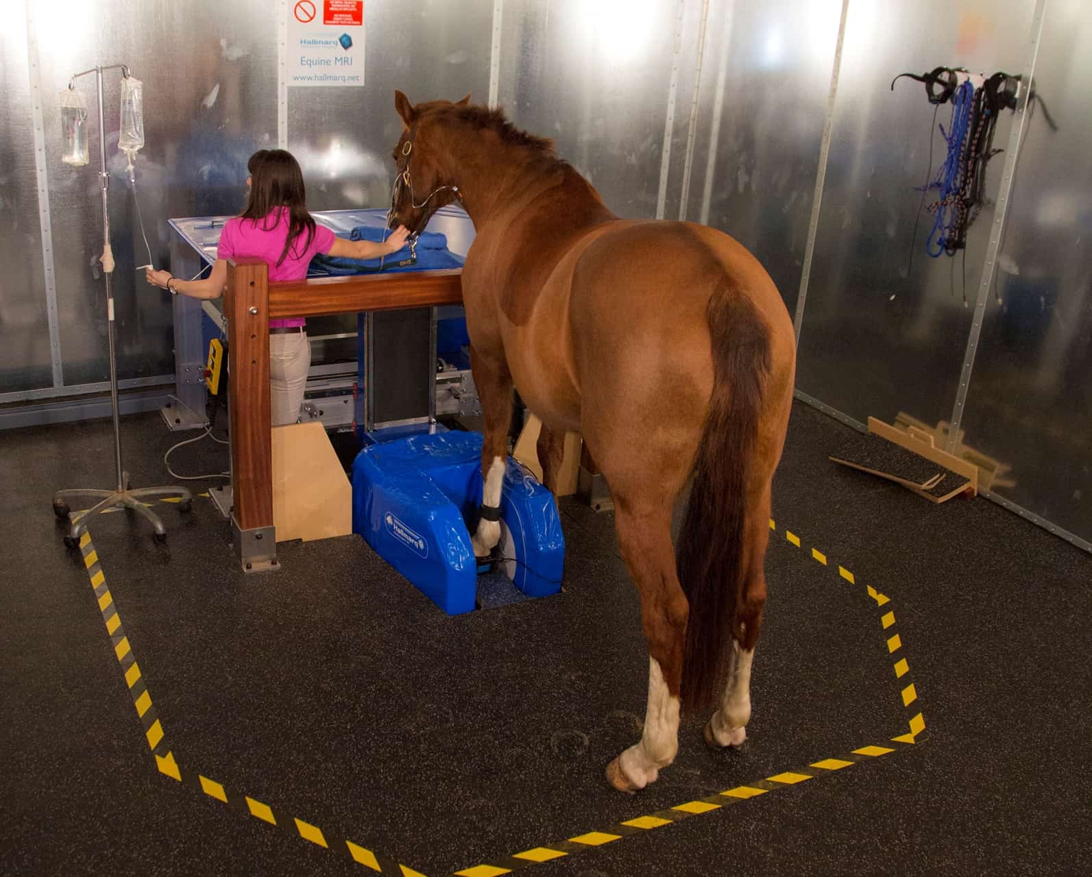
Advances in medical technology aren’t just for people—our equine partners also benefit from an ever-increasing range of sophisticated diagnostic options. Case in point: Horse owners faced with pinpointing lamenesses and other problems now have an arsenal of modern technologies at the ready. Think CT, ultrasound, MRI, and more. So when is a simple radiograph sufficient, and when should you consider bringing in the big guns?
The first thing to keep in mind is that each imaging modality doesn’t exist in a vacuum. Starting with a clinical examination, your veterinary team uses one or more diagnostic techniques to get a better idea of what’s going on inside your horse. Combining close observation and various examination techniques to reach a diagnosis means he or she can pursue a more precise and appropriate treatment plan.
Digital Radiography (X Rays)
Radiographs are the bread-and-butter of diagnostic imaging. Today’s portable machines are easy to use and produce high-resolution images that can be reviewed instantly on a laptop. Because radiographs are reasonably priced and it’s simple to send images electronically for evaluation, veterinarians rely on them for prepurchase and lameness exams
Create a free account with TheHorse.com to view this content.
TheHorse.com is home to thousands of free articles about horse health care. In order to access some of our exclusive free content, you must be signed into TheHorse.com.
Start your free account today!
Already have an account?
and continue reading.
Written by:
Natalie DeFee Mendik, MA
Related Articles
Stay on top of the most recent Horse Health news with


