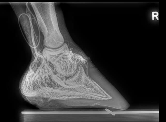
Venograms and Laminitis

—Amy, Houston, Texas
A. In an X ray we can see the coffin bone and other bony structures within the foot capsule, but we can’t really get a good look at the soft-tissue structures, including ligaments and vessels. A venogram is also an X ray, but we place a tourniquet on the leg inject a dye into the vein that feeds the foot. That dye moves into the structures of the foot around the coffin bone and the vessels that feed the laminae (blood flow to the laminae is compromised during laminitis) and, in the image, shows up bright white. It can give us an indication of where there’s leakage of blood in the hoof capsule or maybe limited blood supply
Create a free account with TheHorse.com to view this content.
TheHorse.com is home to thousands of free articles about horse health care. In order to access some of our exclusive free content, you must be signed into TheHorse.com.
Start your free account today!
Already have an account?
and continue reading.
Written by:
Bryan Fraley, DVM
Related Articles
Stay on top of the most recent Horse Health news with


