Horse Hoof Anatomy, Part 1

Understanding the hoof’s inner structures and how they work together
Like the tires and suspension on the truck you drive to the barn, your horse’s hooves take a beating daily. The hoof is cleverly engineered to handle stress, yet hoof problems are unfortunately common. “The foot appears to be the dominant site of lameness in performance horses,” says Stephen O’Grady, DVM, MRCVS, owner of Northern Virginia Equine, in Marshall.
Lameness results from pain in the foot’s inner structures, which are heavily influenced by the outer hoof. O’Grady explains that the hoof is viscoelastic, meaning that it deforms or changes shape dynamically with high stress (expands with weight bearing). It also changes shape or distorts in a more long-term manner with consistent stress (such as contracting over time because of shoes that are too small).
In short, we can cause significant changes in the outer hoof and thereby its inner structures, both for good and for ill, with our management practices. The better we understand how the hoof is made and how it works, the better we can recognize problems brewing and manage the hooves to keep small issues from becoming time-off or retirement-level problems.
In this two-part series we will start with internal hoof anatomy, function, and the most common problems, followed by Part 2 on external anatomy, function, common problems, and management to promote healthy feet. So let’s dive inside, starting with the bones at the center.
Inner Framework (Bones, Ligaments, Tendons)
“The horse’s distal limb is functionally a set of levers and pulleys,” says O’Grady. Moving down the leg from the fetlock, we find three phalangeal bones lined up—the proximal or first phalanx (P1), the middle or second phalanx (P2), and the distal or third phalanx (P3), often called the coffin bone, nearest to the ground and inside the hoof. There’s also the navicular bone just behind the coffin bone that articulates with (unites as a joint) it and P2.
Bones (the levers) provide attachment points for a network of ligaments that hold joints together and tendons that connect muscles to bone (the pulleys). Muscles position the bones to make the horse move. The phalangeal joints allow extension/flexion primarily forward and backward, explains O’Grady. The lower joint possesses a much greater range of motion, while the joint between the coffin and navicular bones allows very little movement.
Two major tendons also help support and move the bones: the extensor tendon that attaches to the top/front of the coffin bone and straightens the leg; and the deep digital flexor tendon that runs down the back of the leg, bending over the navicular bone, attaching on the bottom of the coffin bone, and bending or flexing the leg.
What can go wrong
Arthritis can develop in the hoof’s joints, just as it does in humans’ hands and feet, says Amy Rucker, DVM, owner of Midwest Equine, in Columbia, Mo. Roughening or wearing down of the cartilage that covers the ends of bones at joints and extra bone growth, such as bone spurs, can cause pain and impede the bones’ smooth movement against each other as the horse moves. Ringbone, or excess bone growth at the coffin joint, can develop in response to concussive stress.
Joint function isn’t just about bones, Rucker reminds us. Traumatic injury or stress to the supporting ligaments and/or tendons is another major cause of joint-related lameness. Aside from strains, sprains, and tears, small points of ossification (hardening into bone) of the ligaments where they attach to the bones, called enthesophytes, can also cause lameness.
And, of course, an obvious potential problem with bones is fractures, which can range from small chips to major breaks. The fracture’s severity and location dictates the resulting level of pain and lameness.
Special Bones
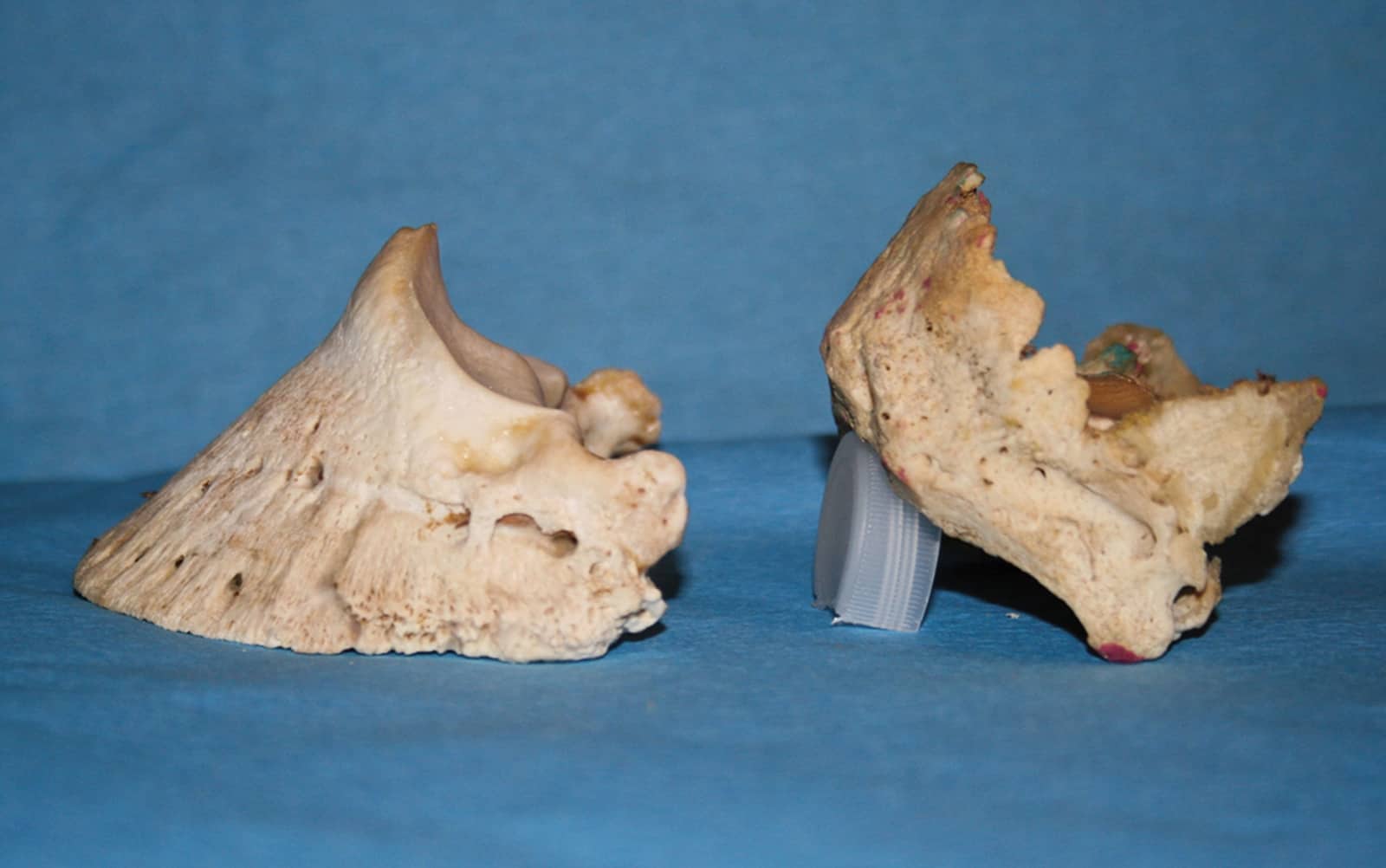
The coffin and navicular bones are functionally quite different from the other bones we’ve described. Along with providing attachment points for tendons and ligaments, the coffin bone has a large, porous surface mirroring the hoof wall angle that’s ideal for soft tissue attachment and blood vessel provision for those tissues. In short, the coffin bone is the outer hoof capsule’s anchor.
This bone also possesses a unique shape (see photo), and among different horses it’s even more unique. The angle between the bottom (solar) surface of the coffin bone and the front of the bone is usually around 50 degrees, says Rucker, but it can be steeper (usually in a club foot) or flatter (usually in a long-toe, low-heeled foot).
What can go wrong with the coffin bone
Like any bone, the coffin bone can fracture and remodel significantly in response to stress. “You will often have loss of the distal rim (toe edge) of the coffin bone if there isn’t much foot or sole mass (for shock absorption and protection),” says Rucker. “This can be secondary to (caused by) poor foot growth, lack of sole thickness/protection, or club foot.”
The palmar angle, between the bottom or wings of the coffin bone and the ground, can be a significant indicator of foot health. For example, a normal palmar angle is slightly positive (heels slightly higher than the toe). But Rucker says an angle higher than 10° suggests the club foot is severe or laminitis has caused instability of the bone within the hoof (and severe laminitis—more on this shortly—can cause coffin bone breakdown starting at the rim and even significant loss of bone mass).
Conversely, a zero or negative palmar angle (heels lower than the toe) suggests that the foot has crushed heels and a compromised digital cushion (more on this coming up, as well).

The navicular bone exists to provide a change in angle for the deep digital flexor tendon running down the back of the leg, says Rucker. Because this tendon is the primary structure that flexes (bends) the entire leg, the navicular bone undergoes significant compressive force.
What can go wrong with the navicular bone
In addition to fractures, the navicular bone is prone to certain injuries often lumped together as navicular syndrome (commonly occurring in low-heeled feet). Rucker notes that edema (fluid buildup) in the bone, often caused by strain and concussion tied to poor conformation or traumatic injury, can result in pain and lameness.
Also, as all bones do, this one can remodel (break down and re-form to change its shape) in response to stress. In the case of the bone’s outer surface, explains Rucker, it might roughen as the old bone breaks down. This in turn can damage the deep digital flexor tendon running over it, and the combination of remodeling and tendon injury can result in the tendon attaching to the remodeled area as it attempts to heal. This limits its function as it can no longer move smoothly across the bone.
Remodeling can also occur in the bone’s interior, but these findings are not yet fully understood.
Collateral Cartilages
The collateral cartilages extend upward from either side of the coffin bone and help the hoof expand with weight bearing.
What can go wrong
Ossification of the collateral cartilages can limit the hoof’s natural expansion and result in some sensitivity to touch via hoof testers, although lameness usually doesn’t occur. Veterinarians see this most commonly in heavy horses such as drafts working on hard streets.
Digital Cushion
The digital cushion does exactly what its name suggests—it lies below the coffin bone (digit) and cushions it against the ground (primarily in the rear of the foot).
What can go wrong
Veterinarians consider the digital cushion to be compromised when a horse has a broken-back hoof-pastern axis and/or long-toe, low-heel conformation. With either issue, the heels are loaded more heavily than they should be and the horse’s weight slowly crushes the cushion to a fraction of its healthy thickness.
“The most common problem I see in internal hoof structures is poor heel structure and inadequate sole depth (due to digital cushion loss),” says O’Grady. “It’s like running on concrete without shoes. If you lose the digital cushion, it doesn’t come back.”
Corium/Laminae
Between the coffin bone and the hoof wall lies the corium, which is soft tissue that includes blood vessels, nerves, and the laminae—interlocking leaflike structures that attach the hoof to the bone. Blood supply to any body part is critical for nourishment (especially when healing from injury or disease), and nerves communicate signals between the feet and the brain and spinal cord.
What can go wrong with blood vessels
Because the hoof wall is mostly rigid, some disease processes can compromise the foot’s blood supply. If a disease or trauma causes swelling in the hoof, the hoof can’t expand to accommodate both the swelling and the blood flow, and swelling generally wins. Without blood flow, tissue and bone can die.
What can go wrong with laminae
Laminitis (also called founder) is disease of the laminae that can range from mild and transient to severe and chronic. The latter cases are degenerative and extremely painful. Its many causes include metabolic problems such as insulin resistance and trauma such as extended work on hard surfaces. At worst, it can cause the laminae to detach from each other and the coffin bone to sink down within the hoof capsule, potentially even punching through the sole.
“Laminitis is the most common problem I see in the inner hoof structures in my practice, and it can often be prevented,” says Rucker. “Most cases I see are caused by insulin resistance or Cushing’s disease, both of which are treatable with diet, medication, and exercise.”
Staying on Top of Things
Rucker recommends having your veterinarian take annual radiographs of your horse’s feet to keep an eye on any brewing problems and to work with your farrier to develop a trimming/shoeing plan.
“Radiographs allow the farrier and veterinarian to assess shoe placement in relation to the stressed areas of the foot and to screen for laminitis,” she explains. “A horse may be ‘sound,’ but not happy. Try to recognize when your horse is drifting past his acceptable range and address the load within the foot, forces of breakover, etc., before you have irreversible damage to the internal structures.”
“It’s important for horse owners to understand the inner hoof structures for your horse’s soundness and to be able to have meaningful discussions with your farrier,” adds O’Grady. “It helps you open an intelligent conversation based on your experiences, not just something you read online.”
In a future article, we’ll look at external hoof anatomy and function and how veterinarians and farriers detect internal problems by evaluating the outer hoof capsule.
Written by:
Christy M. West
Related Articles
Stay on top of the most recent Horse Health news with


