
Measuring Joint Damage
New research is leading veterinarians one step closer to being able to detect the first stages of cartilage damage in joints, which could lead to crippling osteoarthritis. Researchers eventually want to analyze joint fluid or blood samples and assess the concentration of specific biochemical markers thought to be involved in the degradation process of cartilage to predict actual damage i
- Topics: Article
New research is leading veterinarians one step closer to being able to detect the first stages of cartilage damage in joints, which could lead to crippling osteoarthritis. Researchers eventually want to analyze joint fluid or blood samples and assess the concentration of specific biochemical markers thought to be involved in the degradation process of cartilage to predict actual damage in joints.
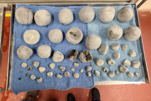
Researchers at Utrecht University in the Netherlands have established a digital imaging technique to quantitatively measure the amount of osteoarthritic damage to a horse's fetlock joint. Harold Brommer, DVM, of the Department of Equine Sciences at Utrecht, said, "Quantification of osteoarthritis is important because the disease is the most common cause of lameness in horses, and the problem is that the initial stages of the disease, starting with cartilage degeneration, are very difficult to diagnose. In the initial stages, there are no radiographic changes because cartilage changes cannot be seen on a radiograph. However, it is very important to detect early cartilage damage in this way because damaged cartilage cannot be fully repaired, making all treatments primarily palliative," explained Brommer. "For that reason, research is focused on prevention nowadays."
Currently, veterinarians must examine the joint arthroscopically (now the golden standard) or anesthetize the horse for magnetic resonance imaging (MRI) to diagnose damage. Scientists around the world have made dramatic advances in the field of osteoarthritis research in recent years, gaining a better understanding of serum and joint fluid markers that might signify cartilage damage. However, the value of these potential markers can only be assessed if their concentrations can be linked to the amount and severity of cartilage damage. So far, no quantitative methods existed to measure this damage reliably. The histological scoring systems that were most often used suffered from the fact that osteoarthritis does not evenly affect the cartilage layer, making samples taken from a certain area not representative for the entire joint.
Scientists in the Utrecht study solved this problem by comparing digital images of the fetlock joint surface before and after staining with Indian ink, a process that has been used in a semi-quantitative way in the past by other researchers to examine damaged joints. Ink particles do not enter intact, undamaged joint areas, but are attracted to damaged cartilage. Studies have shown that the intensity of ink uptake is related to the reduction in the proteoglycan content and surface fibrillation of cartilage, signifying cartilage breakdown. Computer-controlled gray level analysis of the non-stained and India ink-stained cartilage surfaces of the joints and gray scale reference calibration targets was performed, and an increase in the mean pixel value after staining was used to calculate the cartilage degeneration index (CDI)
Create a free account with TheHorse.com to view this content.
TheHorse.com is home to thousands of free articles about horse health care. In order to access some of our exclusive free content, you must be signed into TheHorse.com.
Start your free account today!
Already have an account?
and continue reading.

Related Articles


Stay on top of the most recent Horse Health news with


