
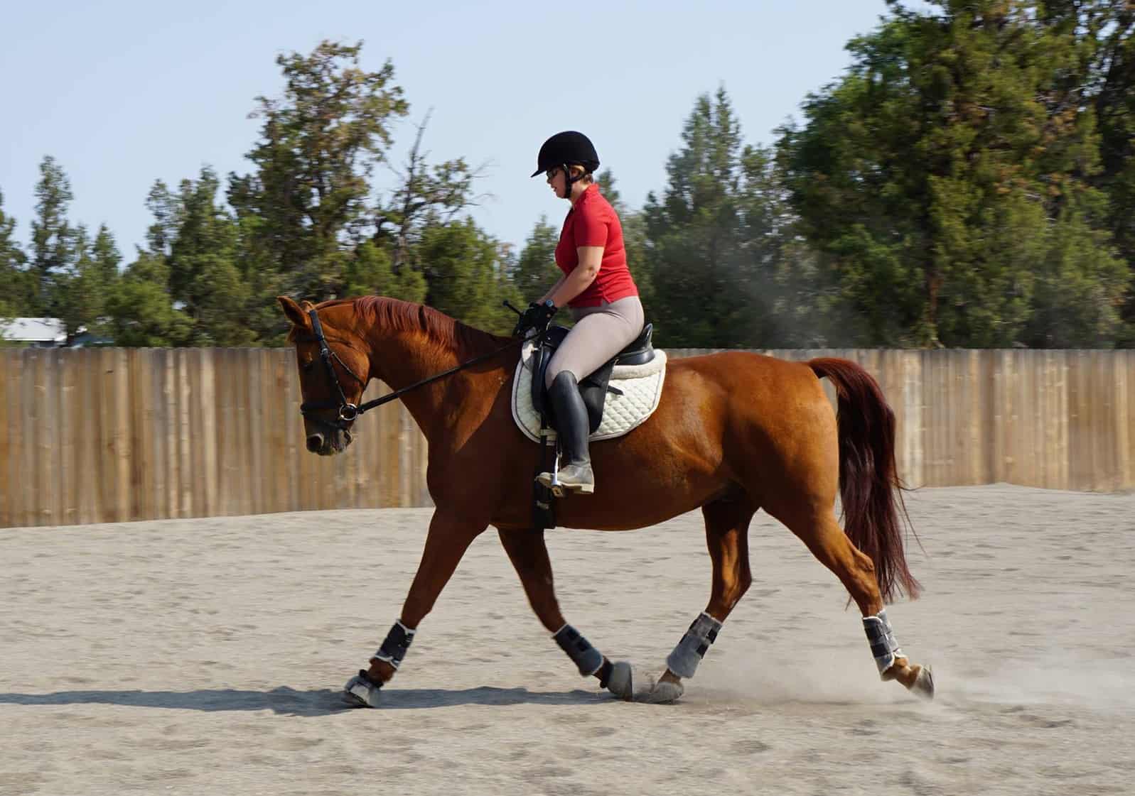
The horse’s heart is well-described in training texts and literature: It is big, giving, trusting, and can push the animal toward great feats of speed and strength. But as much credit as we give horses for their metaphorical hearts, the actual blood-pumping organs generally get far less attention than other bodily systems, said internist Virginia B. Reef, DVM, Dipl. ACVIM, ACVSMR, who specializes in equine cardiology.
As a remedy, the American Association of Equine Practitioners (AAEP) gave the horse’s heart a little love and invited Reef to present the Frank J. Milne State-of-the Art Lecture at its 2018 convention, held Dec. 1-5 in San Francisco, California. Reef, who serves as a professor and section chief in imaging at the University of Pennsylvania School of Veterinary Medicine’s (Penn Vet) New Bolton Center, in Kennett Square, has spent her 40-year career studying the equine cardiovascular system and treating heart disease.
“It’s an area that veterinary schools in the past overlooked—(people believe) horses don’t get cardiac disease or that it’s unimportant,” Reef said. “But there’s more and more recognition that cardiac disease actually is important, and there’s concern from the public and veterinarians about horse safety and sudden cardiac death.”
In Reef’s lecture, “Straight from the Heart: Untangling the Complexities of the Equine Cardiovascular System,” she covered the cardiovascular exam, common cardiac abnormalities, and treatments in-depth.
The Amazing Equine Heart
The horse is an amazing athlete in part because of the efficient way its cardiovascular system pumps blood and, thus, oxygen to the body, Reef said. During exertion, equine cardiac output increases eight to 12 times over resting value, and the horse’s heart rate increases up to eightfold from the typical 32 to 36 beats per minute (or in the mid-20s for an exceptionally fit horse, Reef noted) to a maximum of 240-plus beats per minute.
This, combined with the horse’s large lung capacity and extensive capillary network, allows the horse to consume up to 40 times more oxygen during exercise that it would at rest. “The horse has an impressive set of lungs,” Reef said, emphasizing the important interplay between the cardiovascular and pulmonary (respiratory) systems.
The equine heart weighs an average of 8 pounds and has four chambers: the right and left ventricles and the right and left atria. The mitral, tricuspid, aortic, and pulmonary valves control blood flow from chamber to chamber and into the great vessels (the aorta, which carries oxygenated blood from the left ventricle of the heart through the arteries to the circulatory system, and pulmonary artery, which carries blood from the right ventricle of the heart to the lungs for oxygenation).
A healthy horse’s heart makes up to four sounds per cycle (S1-S4), and Reef noted that each sound is best heard from different locations. When all goes right, blood flows smoothly and efficiently through the heart. When things go wrong, horses can exhibit poor performance and, in some cases, die.
The Basic Cardiac and Prepurchase Exams
Veterinarians might identify cardiac-related issues incidentally during prepurchase exams (PPE), wellness checks, or performance (if the horse is showing clinical signs of heart disease, such as uncharacteristic slowing midrace or knocking of rails during a jumper round).
Reef said the basic cardiac exam starts with a typical physical examination, during which the veterinarian listens to both sides of the heart and all four of its valve areas. “It’s the basic physical exam we all learned in vet school, and the critical part of that exam is the stethoscope,” she said.
The stethoscope allows for “auscultation” which means listening to the sounds the heart, lungs, and other organs make. While examining a healthy horse, the veterinarian should find the heart rate within the normal range; however, Reef noted that anxious or excited horses might have a much higher heart rate than normal.
Next, the veterinarian will palpate the arterial pulses to see if they feel normal. If they’re not, “that’s a clinical clue that will help you differentiate the severity of one of the murmurs (irregular heart sounds) we typically see in middle-aged horses, which is aortic regurgitation (blood flowing backward through the aortic valve),” Reef said.
She follows arterial pulse palpation by looking for a visible pulse in the jugular vein (there shouldn’t be one). Then, during exams at New Bolton Center, the veterinarian takes the horse’s blood pressure using a tail cuff. This cuff works like the arm cuffs used on people to take the horse’s systolic and diastolic measurements (This is similar to the two numbers you see on your own blood pressure measurement: the systolic is the higher top number, indicating the amount of pressure in your arteries during heart muscle contraction, while the diastolic is the lower bottom number, showing the pressure when your heart muscle is between beats).
Finally, the veterinarian should check for healthy mucous membranes: ideally, they’re are pink in color with normal capillary refill times (how long it takes for the gums to return to pink after being pressed with a finger—typically 2 seconds or less). This indicates normal peripheral blood flow (that which passes through the veins and arteries not in the chest or abdomen), Reef said.

Horses with suspected murmurs or arrhythmias (irregular heartbeat) require additional diagnostics. The veterinarian can further investigate using diagnostic tools that allow them to listen to and capture images of the heart, including:
Electrocardiography (ECG) measures the heart’s electrical activity , producing a visual trace on paper or screen, and is used to evaluate the horse’s cardiac rhythm. While equine veterinarians have used ECG for decades, recent technological advances have made its application easier. For example, smartphone-enabled ECG is an inexpensive way to diagnose an arrythmia, Reef said. “You can send that recording to a cardiac specialist to help with the rhythm diagnosis,” she told the veterinary audience.
Additionally, continuous ECG using smartcard recorders offers 24-hour recording. Clinicians can synchronize these recordings with video recordings to correlate abnormal behavior or episodes of collapse or near-collapse with a horse’s cardiac rhythm. Another tool, telemetry (recording and transmitting from a distance), allows clinicians to monitor ECG in real time while the horse is exercising.
Echocardiography (echo) uses sound waves to collect imagery of the moving heart in real time. The addition of Doppler ultrasound to standard echocardiography can also show the speed and direction of blood flow within the heart chambers and through the valves.
Another recent advancement is the four-dimensional echocardiogram, which looks at the three-dimensional images over the fourth dimension of time. “These 4-D images can enhance visualization of congenital (the horse is born with it) cardiac defects and valvular pathologies (disease or damage),” Reef explained.
During a prepurchase exam, abnormalities the veterinarian identifies while listening to the heart might indicate the need for further cardiac imaging and consultation with a specialist. Patient history is important, as well, and the seller should disclose any known heart issues, which could support including an ECG and/or echo as part of the PPE workup.
Accurate diagnosis is important for not only the health and welfare of the horse but also the safety of those who might ride or drive the horse in the future. Specifically, horses with certain cardiac conditions should not be ridden by children, Reef said. A group of international equine cardiac specialists, including Reef, created a consensus statement highlighting the importance of the “informed adult rider” when it comes to horses with certain heart problems that stay in work.
“Riding horses is a risk,” she said. “You can get hurt on a perfectly normal, healthy horse. So, if the adult rider is informed of the risk that there’s a possibility the horse might collapse, then that adult can decide whether or not to ride that horse. A child rider can’t make that decision or the horse’s well-being while it’s being ridden.”
When Things Go Wrong With the Equine Heart
Reef’s presentation included case studies on and sound files of horses with a variety of heart issues seen at Penn Vet, ranging from murmurs to arrythmias to infections. While horses are occasionally born with some of the conditions, other abnormalities are caused by exercise, drugs, infectious disease, toxin exposure, metabolic problems, electrolyte imbalances, and even bites from venomous animals such as snakes. Many conditions, specifically certain murmurs, occur along with or cause additional heart problems, such as irregular heartbeat, and lead to compounding health problems for the horse.
Murmurs—Blood makes sounds as it whooshes through the pumping heart and its vessels. The natural cardiac cycle includes a period of quiet, and a heart murmur is an abnormal audible sound that occurs during that quiet cycle. Murmurs are either congenital or acquired (it develops with age). Veterinarians detect physiological flow murmurs frequently in horses, Reef said, and their job is then to differentiate these functional murmurs from those associated with underlying heart disease. The complete cardiac examination helps the veterinarian diagnose the murmur type and severity, as well as its impact on performance, life expectancy, and horse and rider safety. Vets should perform echocardiographic examinations in all horses with medium to loud systolic murmurs, and any long diastolic murmurs, to determine their cause and significance, Reef said.
Practitioners grade murmurs by intensity or loudness on a six-point scale, with 1 being the least severe and 6 being the most severe. They also described them by:
- Timing;
- Duration;
- Quality (“musical,” for example);
- Point of maximal impulse, or PMI (when the murmur is loudest); and
- “Shape” of the sound.
All of those characteristics can give the veterinarian clues to better understand the murmur, Reef said.
Whether a murmur affects a horse’s performance or life expectancy depends on its cause, severity, and location. For example, aortic regurgitation (AR, which occurs when the aortic valve doesn’t close tightly) is a common finding in middle-aged horses. In most cases, it progresses slowly and doesn’t shorten life expectancy, but in certain instances it can lead to death, she said.
Though Reef described several other types of murmurs that affect horses of different ages and with different athletic demands, she said mitral regurgitation (MR) is the murmur general practice veterinarians encounter most frequently. It’s common in horses of all disciplines, often mild, and doesn’t affect performance or life span unless it occurs at an early age, she said. “Often, it never progresses.”
For moderate heart murmur cases, drugs can extend performance career and life span for low-intensity work. Veterinarians can use ACE inhibitor (angiotensin-converting enzyme inhibitors, which help relax blood vessels) drugs to treat MR and AR; however, Reef noted that the organizing bodies in certain disciplines might prohibit these drugs.
More serious cases have a poorer prognosis and limit a horse from competing in high-intensity, high-risk sports, such as racing or eventing, she said.
“If you have a patient with mitral or aortic regurgitation, you’ll want to assess and document the severity … and we usually recommend an annual exam,” she said. “If you have a horse with a dilated pulmonary artery, that horse is unsafe for anyone to ride because the pulmonary artery isn’t designed to be under high pressure (because the vessel is more prone to rupture). Left atrial enlargement (is) a concern in high-risk sports because it predisposes the horse to atrial fibrillation (AF).”
Arrythmias—Irregular heartbeats have many causes in horses that result in different clinical signs. Horses with AF, for example, might start strong in a race, only to slow halfway to the wire. However, certain atrioventricular blocks occur in fit horses, disappear during exercise, and don’t affect performance, Reef said.
Treatment for arrythmias can include cardioversion, which is a procedure that restores a normal heartbeat either with drug treatment or by shocking the heart with electrodes. While not widely available, transvenous electrical cardioversion (known more commonly as TVEC) “is very successful in horses with AF and no other underlying cardiac disease,” Reef said.

Myocardial disease—Exposure to certain toxins and drugs, as well as hypoxia (lack of oxygen), are a few causes of heart tissue (myocardium) damage. “Most of the time we don’t know for sure what the cause of underlying myocardial disease is,” Reef said.
Specifically, myocardial disease in horses can be related ionophore consumption. Ionophores are antibiotics sometimes added to cattle and chicken feed are toxic to horses (monensin is one example). The death of one or more horses is often the first sign of ionophore exposure, Reef said. Horses that live can suffer long-term negative effects. However, Reef noted that some horses recover and return to active performance careers.
Pericardial disease—Infection of the pericardium (the membrane that encloses the heart) is rare in horses but has been associated with pleuropneumonia and, in a specific outbreak in Kentucky, mare reproductive loss syndrome. Treatment includes pericardial drainage and lavage, which involves removing the fluid and treating the horse with antimicrobial drugs. Early diagnosis and aggressive treatment are necessary for a positive outcome, Reef said.
Congestive heart failure (CHF)—A variety of heart conditions can cause congestive heart failure, which is a weakness of the heart and fluid buildup in the lungs and surrounding body tissues. Because of the interplay between the cardiac and pulmonary system, the lungs and pulmonary artery are often affected, Reef said.
“In the horse, when they get pulmonary edema (fluid swelling), they typically get interstitial edema, and the clinical signs are subtle,” Reef said. “Usually they have a little cough, the respiratory rate is normal at rest but raised maybe a little after exercise; they might have flared nostrils and won’t recover well. These horses are often , and it sounds like their lungs are harsh, so you think, ‘This horse has lower airway disease—it has asthma.’ And many older horses do have some sort of airway disease, as well.”
The result is potential misdiagnosis, Reef said.
Sudden cardiac death (SCD)—This is another risk for horses with certain heart conditions that can result in catastrophic death. It occurs in horses during or shortly after exertion and can result from myriad causes, including internal hemorrhage from ruptured heart vessels. The aorta can rupture into the heart, creating an aortocardiac fistula, a condition that doesn’t immediately cause SCD. These horses are unsafe to ride or drive, Reef said
Take-Home Message
The horse is a superb athlete, thanks in large part to an efficient cardiovascular system. However, historically, these structures haven’t received the amount of attention given to other bodily systems associated with, for example, colic or laminitis. Today, with researchers and teachers like Reef leading the way, veterinarians are learning the importance of cardiac disease, the risk it poses to both horse welfare and rider safety, and treatments that can improve affected horses’ performance and quality of life.