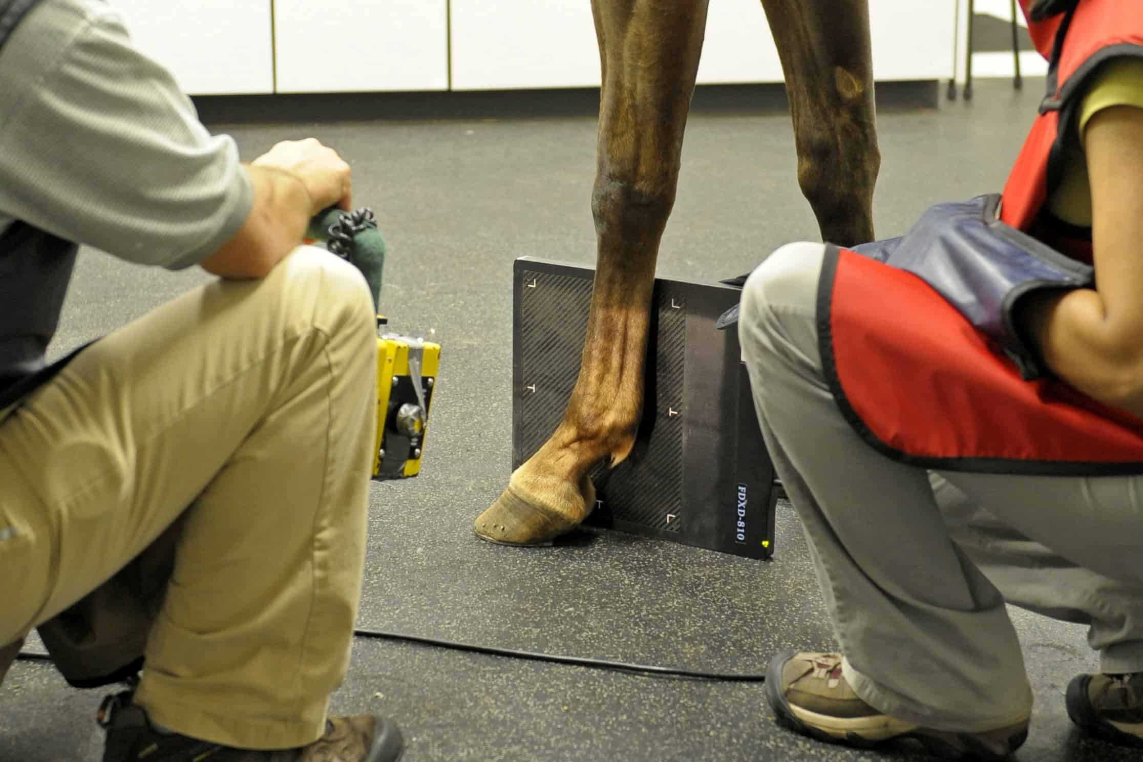
Diagnostic Imaging in Western Horse PPEs: Fifty Shades of Gray

Ultrasound, computed tomography (CT), MRI, and nuclear scintigraphy are finding a place alongside radiography (X rays) when it comes to assessing a horse’s soundness prior to sale. However, with the 50 shades of gray these technologies present, which diagnostic images will be most revealing?
Well, that depends.
During her presentation at the 2019 American Association of Equine Practitioners Convention, held Dec. 7-11 in Denver, Colorado, Florida-based radiologist Natasha Werpy, DVM, Dipl. ACVR, said prepurchase diagnostic imaging in Western performance horses is becoming more complex. Horses across all Western disciplines commonly show wear and tear in their feet, hocks, stifles, and knees. However, each sport tends to manifest its own set of problems
Create a free account with TheHorse.com to view this content.
TheHorse.com is home to thousands of free articles about horse health care. In order to access some of our exclusive free content, you must be signed into TheHorse.com.
Start your free account today!
Already have an account?
and continue reading.
Written by:
Betsy Lynch
Related Articles
Stay on top of the most recent Horse Health news with


