Slow, progressive exercises help improve a horse’s strength and stability when recovering from this neurologic disease

Equine protozoal myeloencephalopathy (EPM) is a neurologic disease caused by the protozoa Sarcocystis neurona or Neospora hughesi and can lead to deficits including ataxia (incoordination), gait abnormalities, and muscle weakness or atrophy.
S. neurona is typically found in the Western Hemisphere, and the definitive host is the opossum. Opossums can get it by scavenging on infected cats, raccoons, and other intermediate hosts. Horses become infected by ingesting feed or water contaminated with opossum droppings. N. hughesi rarely causes EPM in horses, but sporadic cases arise from this organism. Researchers do not yet know what natural hosts can transmit N. hughesi.
Risk Factors for EPM
Researchers estimate 15-90% of horses have been exposed to S. neurona and, therefore, have antibodies in their bloodstream. However, exposure does not always lead to infection—the annual incidence of EPM is less than 1% in the United States. Horses diagnosed with EPM infection are often younger than 4 or older than 13 years old.
Veterinarians see the highest number of EPM cases during the fall, while the disease is much less common during the winter months. Stress, including immune system compromise, intense exercise, transportation, injury, surgery, or foaling can increase a horse’s chances of developing clinical (apparent) EPM.
Diagnosing EPM in Horses
When a horse exhibits clinical signs of EPM, veterinarians must distinguish the disease from lameness or other neurologic conditions such as cervical vertebral stenotic myelopathy (wobbler syndrome), equine
herpesvirus-1 myeloencephalopathy (EHM), rabies, and Lyme disease. Currently, the only definitive way for veterinarians to diagnose EPM is by identifying parasites in the brain or spinal cord during a necropsy.
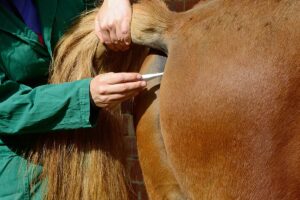
Veterinarians typically diagnose EPM in horses by conducting SAG2, 4/3 ELISA testing, which is performed on serum and cerebrospinal fluid (CSF). They compare the antibody levels in the bloodstream to those in the CSF to see if CSF antibody levels are higher than expected compared to normal diffusion of antibodies from the blood. This would indicate antibody production in the central nervous system. Some practitioners might take cervical radiographs to check for spinal changes that appear more significant than expected with normal aging and could indicate another pathology as the cause of neurologic deficits.
Treating EPM in Horses
Veterinarians have several FDA-approved drugs available to treat horses with EPM: triazines and folate-inhibiting drugs. They also often recommend supplementing with vitamin E.
Researchers have suggested the triazines diclazuril and ponazuril both target the apicoplast organelle in S. neurona, which mammal cells do not contain.
Sulfadiazine and pyrimethamine are folate-inhibiting drugs used in combination to block folate synthesis in S. neurona, which it needs for survival. Researchers have reported vitamin E’s antioxidant properties can help protect the damaged central nervous system from oxidant injury.
With FDA-approved treatment, 57-62% of horses show improvement in clinical signs, but about 10% of horses relapse within one to three years of treatment.
Preventing EPM in Horses
Limiting opossum access to your horse’s feed and water sources remains one of the best ways to lower his risk of S. neurona or N. hughesi infection. Decreasing his stress levels and improving his overall health can also positively impact immune function, possibly lowering his risk of protozoal infection. In some situations your veterinarian might recommend intermittent treatment with low doses of diclazuril to reduce infections.
A Challenging EPM Case
In September 2020 veterinarians at the University of Georgia’s School of Veterinary Medicine, in Athens, admitted George, a then-15-year-old Warmblood gelding used for dressage presenting with moderate neurologic deficits. A few days into his hospital stay, Katie Ellis, DVM, MS, Dipl. ACVSMR, clinical assistant professor, was brought onto the team.
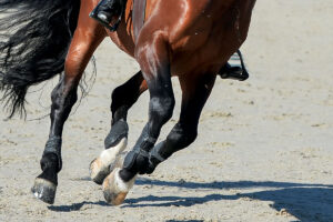
In his case report George’s clinicians described him as bright, alert, and responsive on admission to the hospital. He was very ataxic in the hind limbs as he walked off the trailer and while being turned around in his stall, and in the hospital he dog-sat (a common sign of ataxia).
They also observed that George’s neurologic deficits were more pronounced in the hind limbs, especially on the right, than in the forelimbs. When his vets observed him on tight circles to both the right and left, they reported more significant gait deficits.
George’s clinicians performed cervical radiographs and SAG2, 4/3 ELISA testing to confirm their suspicions of EPM. The cervical radiographs showed only mild changes consistent with his age and work. His CSF analysis revealed high protein levels, which the veterinarians noted can be consistent with EPM infection, and serum titers confirmed their diagnosis.
Treatment and Rehabilitation
George’s veterinarians treated him with ponazuril along with sulfadiazine and pyrimethamine. “These drugs act by two different mechanisms to kill the protozoa and thus stop the parasite from inflicting further damage to the central nervous system,” they wrote in his report. “The gelding’s response to treatment has been positive, and his neurologic clinical signs have continued to improve since starting medication.”
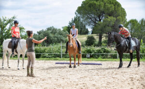
In addition to medical treatment, Ellis and her team developed and completed daily rehabilitation exercises with George to help strengthen his muscles and improve his overall coordination. (See sidebar on page 33.)
Veterinarians employ physiotherapy as one of the first rehabilitation techniques for a neurologic horse, Ellis says.
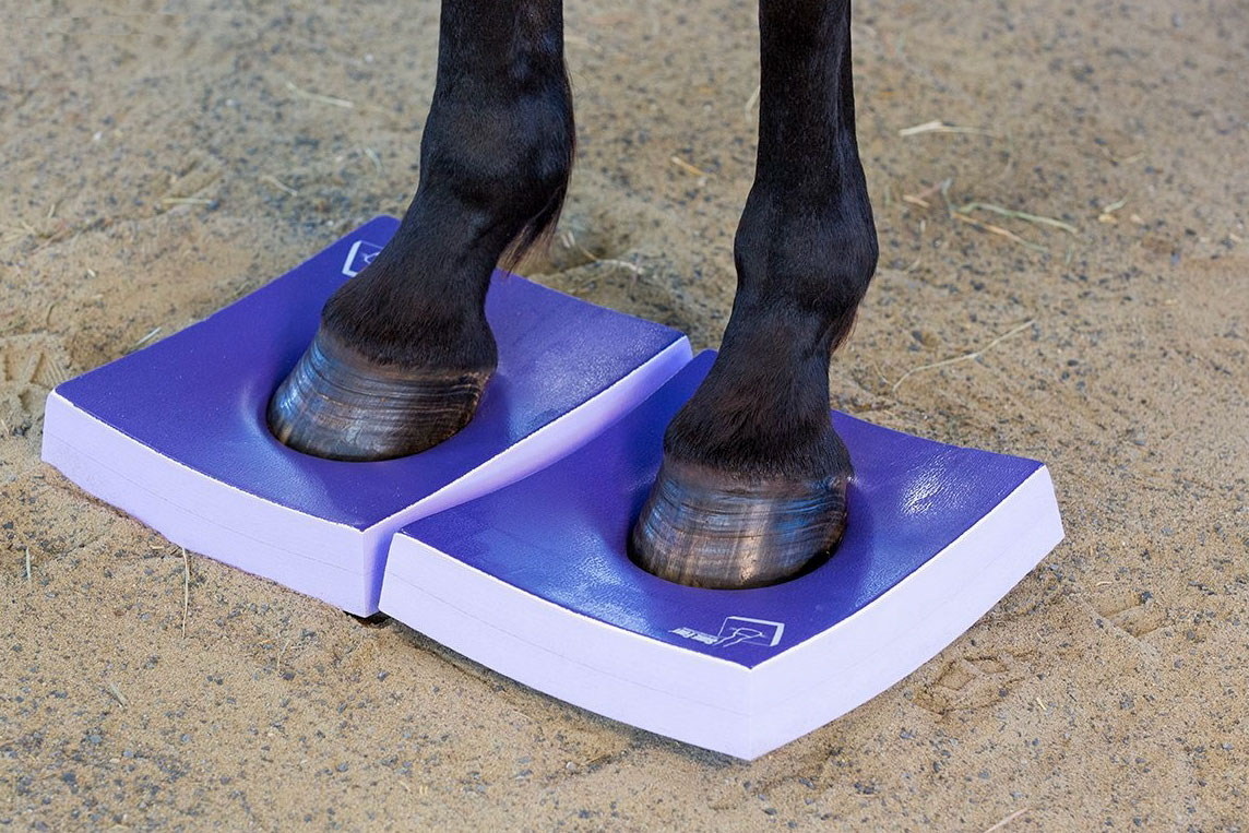
Once George was stable standing on flat ground and able to pick up all four feet, Ellis used proprioceptive balance pads as the next step in improving his coordination and balance. “The horse should start with firm pads and only the front or hind feet on pads before moving to softer pads or placing all four feet on at the same time,” she says. Because this exercise can be very physically challenging for horses, she says she only incorporated it for short periods of time and increased the difficulty slowly based on George’s progress.
After George was stable hand-walking in a straight line, Ellis and her team started incorporating walking him in a serpentine pattern and over surface changes (i.e., from pavement to grass or arena footing).
George’s Future
University clinicians discharged George from the hospital after he underwent 28 days of treatment and rehabilitation. His owner continued his rehabilitation with guidance from and regular check-ins with Ellis. At his last recheck in 2021, George had progressed to light ridden exercise and was continuously improving.
Editor’s note: To protect the horse’s anonymity, we have changed his name in this article.