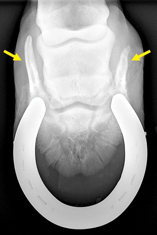
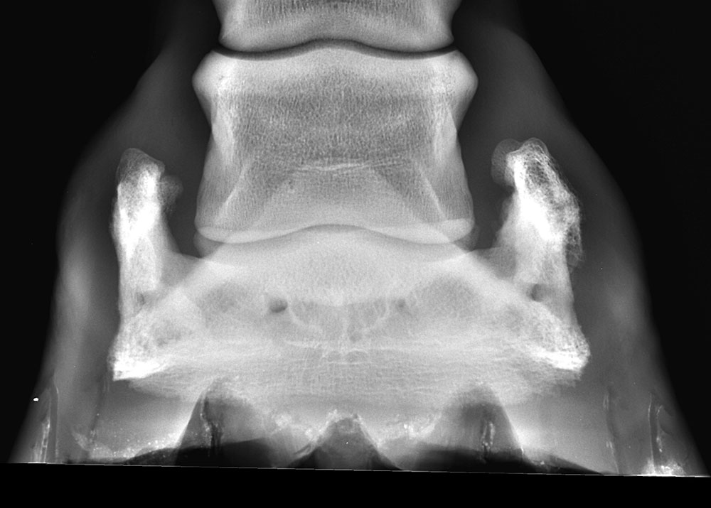
Sidebone refers to ossification of the collateral cartilages of the coffin bone—also known as the distal phalanx (P3) or pedal bone—appearing as upward-extending bony growths. This ossification happens during a process in which the cartilage mineralizes, explains Brian Beasley, DVM, CJF, Dipl. ACVSMR, podiatrist at Grand Prix Equine, a veterinary practice in Newtown, Connecticut.
Essentially, a flexible structure becomes progressively rigid, adds Carlos Carvajal de la Cerda, MVZ, CF, an instructor of equine podiatry at Colorado State University’s Veterinary Teaching Hospital, in Fort Collins. Beasley and Carvajal de la Cerda share their insights into sidebone, from potential causes to best-practice solutions.
Form Follows Function
To better understand sidebone it’s important to review the anatomy and function of the collateral (or ungular) cartilages, says Carvajal de la Cerda, highlighting key points:
- Collateral cartilages can be found above the palmar and plantar processes of the coffin bone (the winglike extensions at the rear of the coffin bone in the forelimbs and hind limbs, respectively). From there, the collateral cartilages extend dorsally to just above the coronary band.
- The collateral cartilages can be seen and/or palpated in the caudal (toward the rear) region of the hoof.
- These cartilaginous structures are highly vascularized (rich in blood supply) and, again, flexible under normal conditions.
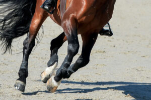
“The importance of the ungular cartilages lies in their nature,” explains Carvajal de la Cerda. “They are designed to aid in shock absorption, along with other structures such as the digital cushion, frog, corium, sole, and hoof wall. When we examine a venogram or dissect a foot, we observe that the back part of the hoof is richly vascularized, forming a perfect hydraulic system to absorb and dissipate impact forces.”
The balance of these structures within the hoof ensures optimal function. “The equine foot possesses a great mechanism to absorb the significant impacts that occur with every stride,” Carvajal de la Cerda says. “The faster a horse moves, the greater the forces on its limbs. Ground reaction forces (transmitted from the ground through the hoof and up through the bony column) also play a role and vary depending on the footing or surface the horse is working on.”
Disruption of this balance could explain why collateral cartilages ossify. “The exact cause of sidebone is not fully understood, but several contributing factors have been proposed,” says Carvajal de la Cerda, including:
- Repeated concussion or trauma to the collateral cartilage
- Conformational imbalances that lead to increased stress on one side of the foot
As for which horses the condition affects most, “sidebone is relatively common in larger breeds such as draft horses, Friesians, and Warmbloods,” says Carvajal de la Cerda. “It may also occur in horses with angular limb deformities. While it is more frequently seen in older horses, young horses are not excluded.”
Diagnosis and Next Steps
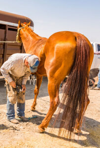
“In my practice, sidebone is most often an incidental finding discovered during radiographic examinations performed for other reasons,” says Carvajal de la Cerda
This story requires a subscription to The Horse magazine.
Current magazine subscribers can click here to and continue reading.
Subscribe now and gain unlimited access to premium content.
Subscribe NowWe at The Horse work to provide you with the latest and most reliable news and information on equine health, care, management, and welfare through our magazine and TheHorse.com. Our explanatory journalism provides an understandable resource on important and sometimes complex health issues. Your subscription will help The Horse continue to offer this vital resource to horse owners of all breeds, disciplines, and experience levels.