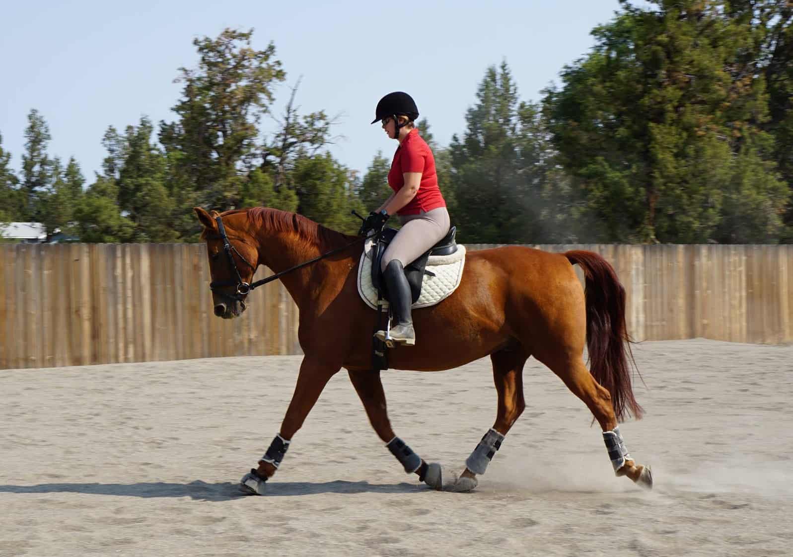
12 Facts About Diagnosing Distal Leg Lameness in Horses
- Posted by Michelle Anderson
Share:

Today’s veterinarians have imaging technologies available to them that have changed and improved the way they evaluate, diagnose, and ultimately treat equine lameness in the lower leg and foot.
Here’s a look at 12 facts and resources from The Horse related to diagnostic modalities for the distal limb. Don’t forget to watch Dr. Brendan Furlong’s presentation, “From Hoof Testers to MRI,” which is part of our Vet on Demand Lecture series organized in partnership with the University of Kentucky’s Gluck Equine Research Center.
Share

Written by:
Michelle Anderson
Michelle Anderson is the former digital managing editor at The Horse. A lifelong horse owner, Anderson competes in dressage and enjoys trail riding. She’s a Washington State University graduate and holds a bachelor’s degree in communications with a minor in business administration and extensive coursework in animal sciences. She has worked in equine publishing since 1998. She currently lives with her husband on a small horse property in Central Oregon.
Related Articles
Stay on top of the most recent Horse Health news with


