Identifying Sacroiliac Joint Pain
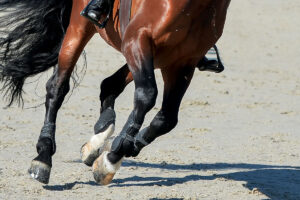
A diagnosis of sacroiliac joint (SI) pain tends to be one of exclusion, after other causes of hind limb lameness have been ruled out. To simplify the diagnostic process, researchers in the United Kingdom conducted a study involving 74 horses suspected of having SI pain. The objective was to use both nuclear bone scan (scintigraphy) and a local anesthetic block to positively identify abnormal
- Topics: Article
A diagnosis of sacroiliac joint (SI) pain tends to be one of exclusion, after other causes of hind limb lameness have been ruled out. To simplify the diagnostic process, researchers in the United Kingdom conducted a study involving 74 horses suspected of having SI pain. The objective was to use both nuclear bone scan (scintigraphy) and a local anesthetic block to positively identify abnormal SI regions. In addition, age, sex, breed, work discipline, size, and conformation were examined to see what type of horses are more susceptible to SI problems.
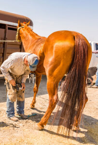
Results indicated that dressage and show jumper horses are at increased risk of SI pain, but only at higher levels of performance. Older, taller, heavier horses are also at increased risk, with Warmbloods representing 51% of cases. With this general physical description in hand, Sue Dyson, FRCVS, of the Center for Equine Studies, Animal Health Trust, Newmarket, United Kingdom, agrees that it’s likely SI pain is a wear-and-tear problem, insidious in onset and chronic in nature. Other results indicated no correlation of SI pain with sex or conformation, and contrary to the long-held belief, asymmetry between the tubera sacrale (top of the pelvis) of affected horses was rare. However, thoracolumbar epaxial muscle atrophy (wasting) and hindquarter muscle asymmetry was common.
All 74 horses had a nuclear bone scan performed, and 73 had a positive result. Thirty-four horses had a local anesthetic block performed by Dyson, and all had profound improvement in gait after 15 minutes. Dyson reminds us that the SI joint is a complex region, and pain can emanate from several places within it. “I think that there can be pain coming from the supporting ligaments and/or the joint. Separation is difficult because the local anesthetic solution can obviously spread from where you put it.” If the block provides virtually complete relief, the diagnosis of SI pain is made. Dyson adds that horses tolerate the 18-gauge spinal needle used for the anesthetic block quite well. Despite this, in her opinion, it is unlikely that local anesthetic infiltration will find use as a therapy for SI pain. Dyson’s other research interests include magnetic resonance imaging (MRI) of the equine foot, demonstrating that a wide variety of lesions can be identified with MRI that are often missed with radiographs or ultrasound. Visit www.aht.org.uk to learn more.
Dyson, S.; Murray, R. Equine Veterinary Journal. 35 (3), 240-245, 2003 TheHorse.com is home to thousands of free articles about horse health care. In order to access some of our exclusive free content, you must be signed into TheHorse.com. Already have an account?Create a free account with TheHorse.com to view this content.
Start your free account today!
and continue reading.
Related Articles


Stay on top of the most recent Horse Health news with