MRI Can Help Diagnose Early Stage PPID
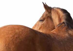
Think “Cushing’s disease,” and a shaggy-looking senior horse might come to mind. This typical image, however, represents moderate to severe pituitary pars intermedia dysfunction (PPID), which can be tough to treat. Catching PPID in its early, more treatable stages would be ideal, but these horses appear much more “normal,” as do their endocrine testing results. One veterinarian has been using MRI to catch these early cases.
At the 2015 American Association of Equine Practitioners Convention, held Dec. 5-9 in Las Vegas, Hal Schott II, DVM, PhD, Dipl. ACVIM, professor of large animal medicine at Michigan State University, described how he and his colleagues confirmed MRI’s usefulness for identifying telltale changes within horses’ pars intermedia (located in the pituitary gland), which are responsible for the development of the debilitating condition.
Previously, Schott’s team had used computed tomography (CT) scans to evaluate PPID-affected and control horses. While they could detect an increase in overall size of the pituitary gland using CT, they confirmed that the anatomic detail within the pituitary gland on CT scans was poor.
So Schott turned to MRI to better define the pituitary gland’s anatomic pathology and develop a grading system score of 1 to 5, with 5 being most severe. Over time, in affected horses, the pituitary gland’s intermediate lobe enlarges and then forms 5-mm or smaller microadenomas (benign tumors) and eventually larger macroadenomas, eventually leading to end-stage disease
Create a free account with TheHorse.com to view this content.
TheHorse.com is home to thousands of free articles about horse health care. In order to access some of our exclusive free content, you must be signed into TheHorse.com.
Start your free account today!
Already have an account?
and continue reading.

Related Articles


Stay on top of the most recent Horse Health news with