Sport Horse Medicine Focus of AAEP’s 2017 Resort Symposium
Lecture topics will include neurologic and orthopedic problems, imaging techniques, and treatment and rehab options.

Lecture topics will include neurologic and orthopedic problems, imaging techniques, and treatment and rehab options.
Dr. Kathryn Wulster is a radiologist and assistant professor of diagnostic imaging at Penn Vet.
Drs. Tim Parkin and Sarah Plevin describe studies focused on predicting injury before it occurs.
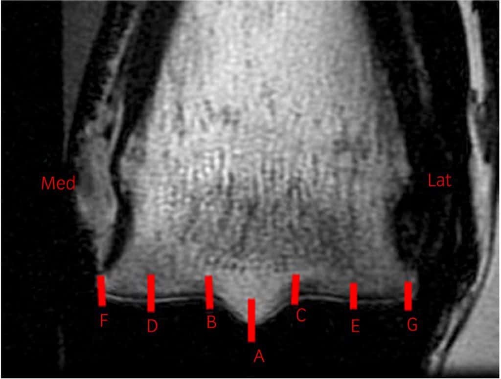
Researchers found that standing MRI is effective for viewing the difficult-to-image ligaments in the horse’s lower legs.
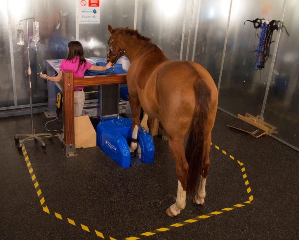
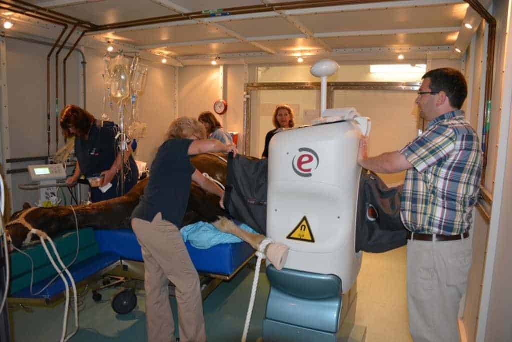
Dr. Kathryn Wulster will use advanced imaging systems, including MRI, CT, and robotics-controlled imaging.

An accurate diagnosis with MRI can play a significant role in a horse’s long-term return to activity after DDFT injury.

Researchers found that “an unexpectedly large number of normal horses” had temporomandibular joint (TMJ) variations.

Frequent findings included osteoarthritic changes, sclerosis, and mild bone spurs, among others.

MRI can identify telltale changes in horses’ pars intermedia, which are responsible for the debilitating condition.

Improved diagnostics and more promising treatments are putting many foot-sore horses back to work.

The horse’s lower limb is subject to a multitude of injuries that can baffle even the most veteran veterinarians.

Poor performers can be a diagnostic challenge. Here’s what veterinarians will look for when examining these horses.
The new MRI replaces a previous system at New Bolton Center, installed in 2005 and used until recently.
Cornell Ruffian Equine Specialists, located next to Belmont Park, celebrated its first year in business on June 25.
Are CT, MRI, and X ray clear as mud? Learn about the appropriate uses for these imaging modalities and more.
The MRI unit adds to the school’s imaging modalities, including a CT, radiography, nuclear scintigraphy, and more.


