DDFT Injury, MRI, and Medical Treatments

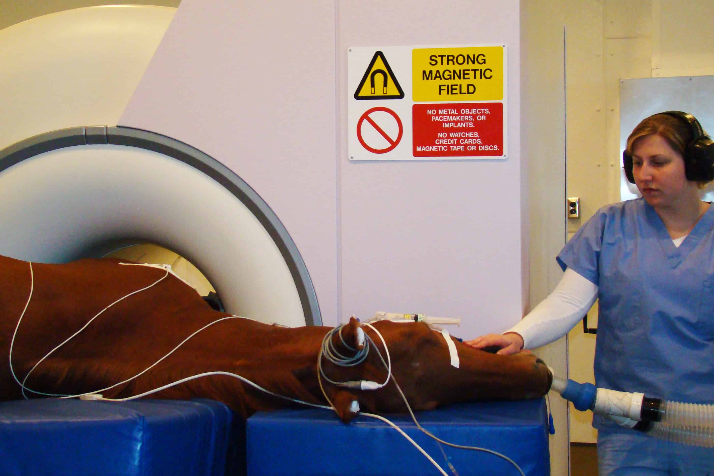
In a recent study, Dylan Lutter, DVM, MS, Dipl. ACVS, and colleagues confirmed that MRI technology more accurately identifies the specific location of the DDFT injury than radiographs, which allows for more targeted treatments. Lutter, a clinical assistant professor of large animal emergency surgery at Kansas State University’s College of Veterinary Medicine, also used MRI to categorize the location and type of DDFT injury while looking for an association with prognosis.
Without MRI results, horses with heel pain are sometimes diagnosed (or misdiagnosed, as the case might be) with “navicular syndrome” and treated without rest, Lutter said. However, his study, which specifically focused on a treatment combination prescribed based on MRI findings, including intrasynovial (used in the joint structure) corticosteroid injection along with six months of rest and rehabilitation—yielded a longer duration of return to use compared to horses that were injected, but not rested.
“The combined steroid and rehabilitation group had a significantly longer duration of return primarily because the rehabilitation program allowed for the tendon to heal and remodel more appropriately than those horses which were injected with corticosteroids and immediately returned to work,” Lutter said.
Corticosteroid injections alone did allow some horses to return to use in the short term. The benefit of combining rest and corticosteroid injection remains unclear, he said.
“The steroids may also have played a role in the improved outcomes,” Lutter explained. “I hope to investigate whether this is the case in the future.”
Lutter suggested that owners of horses with lameness localized in the foot should consider an MRI to either diagnose or rule out a soft tissue injury that could require a targeted treatment plan which might include extensive rest and rehabilitation.
“If an MRI is not possible, it would be reasonable to consider putting your horse through a rehabilitation program as long as there are no severe radiographic changes to the navicular bone,” he said.
The study, “Medical treatment of horses with deep digital flexor tendon injuries diagnosed with high-field-strength magnetic resonance imaging: 118 cases (2000-2010),” was published in the Journal of the American Veterinary Medical Association.
Written by:
Katie Navarra
Related Articles
Stay on top of the most recent Horse Health news with


