duPont Scott Equine Medical Center Gets High-Def CT Scanner
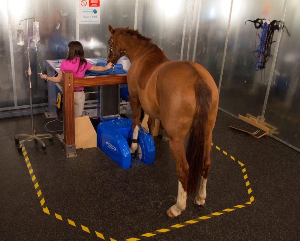
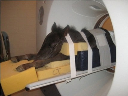
The new unit allows veterinarians to perform high-definition CT scans on standing or recumbent (lying down) horses.

The new unit allows veterinarians to perform high-definition CT scans on standing or recumbent (lying down) horses.
Dr. Kathryn Wulster is a radiologist and assistant professor of diagnostic imaging at Penn Vet.
Dr. Kathryn Wulster will use advanced imaging systems, including MRI, CT, and robotics-controlled imaging.

The new scanner is wider than the standard size, which should allow most of a horse’s neck to be examined.
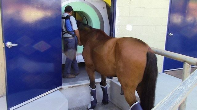
The new CT unit’s gantry is 85 cm in diameter to accommodate large equine body parts, such as the neck.
The four-robot system can perform multiple modalities and will be used in conjunction with a high-speed treadmill.

The horse’s lower limb is subject to a multitude of injuries that can baffle even the most veteran veterinarians.

Poor performers can be a diagnostic challenge. Here’s what veterinarians will look for when examining these horses.
Cornell Ruffian Equine Specialists, located next to Belmont Park, celebrated its first year in business on June 25.
Are CT, MRI, and X ray clear as mud? Learn about the appropriate uses for these imaging modalities and more.
Of the 375 respondents, 201 (54%) said the most recent imaging modality used on their horses was radiography.

Studies covered stem cells, nerve blocks, respiratory issues, joint supplements, kissing spines treatments, and more.
Murdoch University’s new CT scanner is large enough to accommodate horses, as well as smaller animals.
Veterinarians are taking a page from human medicine books by using CT to evaluate pulmonary disease in foals.

One veterinarian believes CT offers a better option for imaging some lower limb fractures than radiography.

Researchers hope to more fully characterize alterations at the microscopic level in diseased navicular bones.