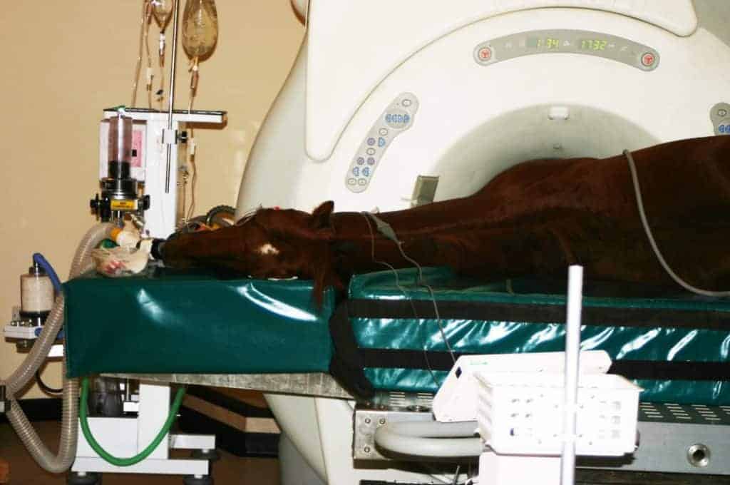
Standing MRI: Use in Diagnosing Equine Lameness
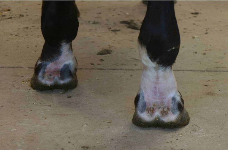
Find out why standing MRI is useful for identifying complex issues in horse hooves and limbs.

Find out why standing MRI is useful for identifying complex issues in horse hooves and limbs.

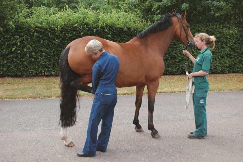
Veterinarians have tools to help them make educated judgments about lamenesses, their causes, and prognoses.
The Animal Health Trust orthopedics team performs cutting-edge research of equine anatomy and function.

One vet said multiple abnormalities could contribute to hoof lameness, rather than just one problem.

Dr. Monty McInturff of Tennessee Equine Hospital shares the most important things he thinks horse owners should know about magnetic resonance imaging (MRI) as a diagnostic tool.
Researchers say MRI is invaluable for identifying suspensory ligament lesions and sesamoid bone damage.

A team evaluated a possible association between distal border fragments of the navicular bone and lameness.
Using both MRI and scintigraphy could be beneficial when diagnosing suspensory-ligament-related injuries

MRI could detect bone changes indicating a horse is at risk for catastrophic fractures before accidents occur.
MRI might offer evidence of laminitic changes in a horse’s hoof before the disease is otherwise identified.

A mobile, high-field unit will be available at the Veterinary Medical Center at least once monthly.
The presentation, “Latest Technologies in Diagnosing Equine Lameness,” was led by Kent Allen, DVM.
Each imaging option, such as MRI, radiography and more, serves a unique role in equine lameness diagnosis.

Researchers recently examined whether diagnostic anesthesia could skew the results of equine foot MRIs.
Researchers found that MRI more accurately identified compression in CSM horses compared to radiographs.
One researcher noted that some defects not visible on radiographs (X rays) were easily identified via MRI.