Accelerating Medical Progress on Equine Lameness
One researcher describes the promising progress that’s been made in lameness diagnosis and treatment.

One researcher describes the promising progress that’s been made in lameness diagnosis and treatment.

Vets can glean crucial information by evaluating horses with performance issues in hand, on the longe, and under saddle.

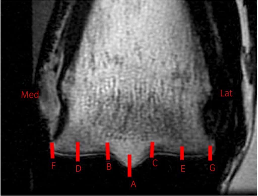
Researchers found that MRI images of bone thickness could provide critical information about fracture risk.

Veterinarians discuss how they diagnose and treat injuries to the collateral ligament, DDFT, navicular bursa, and more.

Tweets and take-homes from sessions on biosecurity, equine collapse, MRI, industry issues, and more!

Find out how veterinarians and farriers rely on imaging to evaluate the horse’s hoof.
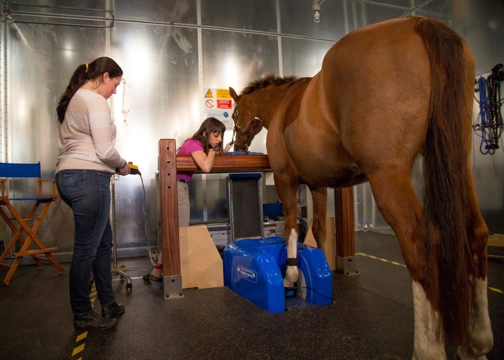
Take a look at the evolution of diagnosing distal limb lameness in sport horse practice with Dr. Brendan Furlong.

Veterinarians can keep diagnostic costs lower by using as much information from routine tests as possible.

Veterinarians have begun research, using the scanner in a clinical trial on client-owned horses.
The new unit allows veterinarians to perform high-definition CT scans on standing or recumbent (lying down) horses.

A look at noninvasive high-tech therapies–from lasers to ceramic-thread blankets–and how they might help horses heal.
Lecture topics will include neurologic and orthopedic problems, imaging techniques, and treatment and rehab options.
Dr. Kathryn Wulster is a radiologist and assistant professor of diagnostic imaging at Penn Vet.
Drs. Tim Parkin and Sarah Plevin describe studies focused on predicting injury before it occurs.
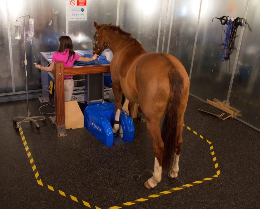
Researchers found that standing MRI is effective for viewing the difficult-to-image ligaments in the horse’s lower legs.
Dr. Kathryn Wulster will use advanced imaging systems, including MRI, CT, and robotics-controlled imaging.