From baseline images to diagnosing complex cases, here’s why X rays should be key components of your horse’s hoof care plan

Radiographs have revolutionized medicine, veterinary or otherwise. In a little over a century, we have gained the ability to look inside a patient’s body and evaluate not just bony structures but also organs and soft tissues.
Veterinarians use radiology in their day-to-day practice. As an equine practitioner, I take radiographs on a very regular basis, with X rays of the hoof being the most common. In this article I’ll describe why hoof radiographs are important, how to use them, and the role they play in helping your horse feel and move better.
What’s in a Radiograph?
The outer part of the hoof capsule is made up of dense keratin, the same material that forms your fingernails. Inside you’ll find primarily the third phalanx (also known as the coffin or pedal bone) and some soft tissue structures. The coffin bone is attached to the hoof capsule by a network of laminae, a scaffolding of soft tissue between the wall and bone.
Radiographs of the hoof also include the second and first phalanx (the short and long pastern bone, respectively). If the horse is on the smaller side, you might see the fetlock joint on the same image.
Betsy Lordan, DVM, CJF, TE, a veterinary podiatrist with SRH Veterinary Services, in Ipswich, Massachusetts, explains the usefulness of hoof radiographs for diagnosing issues and monitoring anatomy: “While we can determine a lot about horses looking at their gross conformation, things are not always what they seem. Using radiographs, we can improve upon those clinical exam findings and fine-tune details which are not as easy to manipulate on the outside of the hoof capsule. Some parameters we evaluate include bony column alignment, sole depth, palmar angle (the angle the bottom of the coffin bone makes with the ground), and evidence of diseases such as arthritis or navicular syndrome (podotrochlosis).”
Baseline Hoof Radiographs
More often than not, veterinarians recommend hoof radiographs because they have narrowed the cause of a clinical lameness to the foot, typically using diagnostic analgesia, or nerve blocks. They use radiographs to evaluate the bony column for pathology (disease or damage). Sometimes, however, clients ask for a baseline set of radiographs on a sound horse, often during a prepurchase exam or on a newly purchased mount.
“Baseline radiographs are very helpful because the hoof is not a static structure—it will morph and change over time and adapt to a variety of circumstances,” says Lordan. “If you’re lucky, you will buy a horse that never has a foot or lameness problem, but the odds are slim.”
Horses can have individual anatomic differences that might have varying clinical significance. Having a good set of baseline radiographs when the horse is sound can help your veterinarian distinguish whether those variations are relevant to a clinical condition, she says. They can also help your veterinarian track the progression of a condition and provide prognostic information.
“I’ve worked on many a white line disease case where the horse was completely sound, and severe hoof separation and capsular rotation were detected incidentally on radiographs,” Lordan says. “Those are the types of cases where the average person would never know there was a problem until the horse was in serious trouble.”
Cases That Warrant Radiographs
Veterinarians use radiographs to adequately diagnose a wide range of clinical cases, including:
Podotrochlosis
Also known as caudal heel pain, podotrochlosis is a chronic condition, usually a bilateral (affecting both sides) lameness of the forelimbs. Kendra Fake, VMD, associate veterinarian at Willow Creek Equine Veterinary Services, in Reading, Pennsylvania, says evidence of podotrochlosis is one of the biggest causes of lameness she finds when assessing hoof radiographs.
Affected horses can have heel pain in response to hoof testers, a choppy gait with their forelimbs, and a noticeable head bob on a circle. “A lameness that blocks to the heel, combined with radiographs that show degenerative navicular (bone) lesions, allows the veterinarian to make recommendations,” says Fake, which can range from corrective shoeing, shock wave therapy, pain management drugs, and joint therapies to further diagnostics such as MRI.
Osteoarthritis
While it can happen in any joint, osteoarthritis is a common finding in horses’ distal (lower) limbs. “The development of osteoarthritis between the coffin bone and the short pastern (the coffin joint) is colloquially known as ‘low ringbone,’ ” says Fake. Then there’s high ringbone, which occurs at the union of the short and long pastern bones, known as the pastern joint. Veterinarians take hoof radiographs to look for arthritis, sclerosis, and other bony changes in the affected joints before recommending treatment protocols, including joint therapies and systemic medications such as phenylbutazone (Bute) and firocoxib (Equioxx).
Subsolar abscess
Most owners have probably managed these localized bacterial infections in the sole of the hoof at some point. The bacteria produce gas that builds pressure in the capsule. This causes the horse immense pain that can easily be confused with a fractured limb.
Usually, these cases do not require diagnostic imaging. However, if a significant amount of time passes with no respite for the horse, the veterinarian might recommend radiographs. While abscesses aren’t always visible on X ray, gas pockets and tracts—formed as the hoof tissues try to relieve the pressure—often are. Your veterinarian can then go in and try to follow these tracts to the abscess, providing drainage and giving the horse instant relief. Radiographs can help ensure nothing more sinister (such as septic pedal osteitis, or infection of the coffin bone) is going on in the hoof.
Keratoma
Uncommonly, horses can develop keratomas. “These are benign tumors coming from the keratin-producing layer of the hoof wall that can cause frequent abscesses and lameness,” says Fake. Generally, horses with keratomas develop several subsolar abscesses in the same spot within a few months. Radiographs can reveal the presence of a keratoma in the body of the coffin bone.
“While sometimes bulging of the hoof wall or distortion of the white line may be seen on visual examination, radiographs that show resorption (breakdown) of the coffin bone where the tumor is invading can be critical in this diagnosis,” she adds. “Once the problem is identified, we can direct the horse toward surgical debridement of the tumor and resolution of the lameness.”
Laminitis
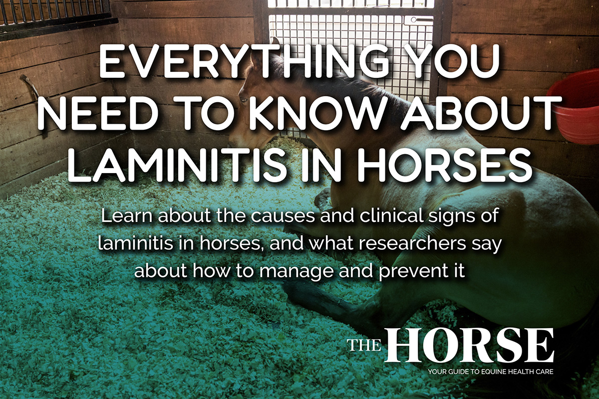
Laminitis is the inflammation of the laminae that attach the coffin bone to the hoof capsule. It can be an incredibly painful and debilitating condition for the affected equid, with chronic inflammation leading to rotation of the coffin bone within the dorsal hoof capsule—sometimes penetrating the sole—or even sinking of the entire bony column.
When laminitis causes coffin bone displacement and/or becomes chronic disease, it’s also known as founder. Determining why a horse has developed laminitis is a priority because several conditions can incite it, and a veterinarian should be involved at the first sign of foot pain. Farriery is very important in keeping laminitic horses comfortable, either with trimming or shoeing.
“The horse’s clinical level of pain is not always correlated to what is going on in the foot,” says Lordan. “Radiographs allow you to assess the degree of the problem and monitor for further deterioration or improved hoof growth in the healing stages. Horses that have been affected by laminitis may continue to show clinical discomfort during the hoof regrowth process, and radiographs help us distinguish that from disease progression.”
A veterinarian’s radiographs are invaluable to the farrier caring for the affected horse’s hooves. “A laminitic horse is a serious situation no matter the age of the horse or cause of the laminitis,” says Tyler Fortier, a farrier servicing New Hampshire and Southern Maine. “Radiographs are critical here. They are basically a road map for how to approach shoeing or not shoeing the horse. It shows a lot of information about what’s going on with the hoof, such as sole depth and the amount of rotation, that’s very valuable in treating a foundered horse.”
Radiograph-Assisted Podiatry
Farriers use hoof radiographs regularly to better care for their patients’ feet. Farriers are often the first line of defense against hoof issues and are frequently on the ground faster than most veterinarians.
“I think radiographs are extremely helpful for me as a farrier,” says Fortier. “My job is to line up the horse’s bones inside the hoof capsule and balance their hooves to support their conformation. What we see from the outside isn’t always correct on the inside. I might feel that my trim is really good, but the radiographs may say something different.”
While some vets and farriers work independently, briefing one another over phone or email, simultaneous visits can benefit case progress greatly. Besides allowing the opportunity to correlate the trim’s gross appearance with on-screen images, Lordan says taking radiographs during shoeing “provides an additional comfort level for the farrier who is being asked to remove a lot of hoof or do something outside their comfort zone. They can gauge their safety window of short vs. too short much more easily. Using radiographs while shoeing allows us to adjust things by degrees, check our work, and make further adjustments as necessary. Taking a set of radiographs gives your farrier the ability to do their best work.”
“Working with a vet as a farrier gives us greater knowledge of the horse we are working on,” adds Fortier. “A good relationship between farrier and vet is when they can work on a lameness together, come up with a common goal, and be confident and respectful to bounce ideas off one another, because not every shoeing script works for every horse.”
Take-Home Message
Hoof health is vital to the horse’s overall health. Hoof radiographs are incredibly useful tools that allow both veterinarians and farriers to do their jobs properly and best treat the horse.