Permanent Tracheostomy Safe and Effective in Select Horses
Creating a permanent hole in the windpipe of horses through a tracheostomy might be the treatment of choice.

Creating a permanent hole in the windpipe of horses through a tracheostomy might be the treatment of choice.
Arthroscopic surgery, developed in the horse in the 1970s, is the keyhole technique by which surgery is performed on equine joints for traumatic injury, fractures within joints, soft tissue injury, and abnormal joint development in young horses,
Cataracts have been found to be heritable in Belgians, Morgans, Thoroughbreds, Rocky Mountain Horses, and Quarter Horses. In other instances, cataracts can develop secondary to trauma or due to chronic inflammation from uveitis (moon blindness).
When it comes to artificial insemination in horses, the site of deposition might have a big impact on the procedure’s outcome. Placing semen directly into the uterine horn containing the ready follicle could allow breeders to use far less
Veterinarians who attended presentations at the event came away with 18 hours of continuing education credit and new knowledge of current topics in equine reproduction and lameness.
Incisional hernias (protrusion of abdominal contents through a gap in an incision beneath the skin) occur in up to 17% of horses receiving abdominal surgery, reported Gal Kelmer, DVM, MS, clinical assistant professor at the University of
The high degree of detail seen with MRI has made it possible for veterinarians to find equine injuries they’ve never seen before. One example of this–MRI evaluation of desmitis in the oblique and straight distal sesamoidean ligaments–was discussed.
Utilizing an arthroscope–a slender instrument for visualizing the environment inside joints–can be a good move when radiographs fail to elucidate the bony cause of a horse’s lameness. Dean Richardson, DVM, Dipl. ACVS, head of surgery at the
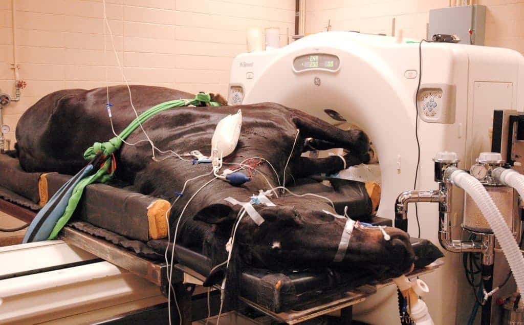
Not all MRI units are created equal. Learn about the differences in MRI units.
The chronically laminitic horse is often a very tough case to manage because displacement of the coffin bone within the foot leads to a lot of pain and damage, in addition to the damage that allowed the displacement in the first place.
Pain originating in the upper cannon bone area, just below the knee or hock, is common in all types of equine athletes. However, it can be difficult to determine exactly what structure is injured; some injuries can only be seen with high-field MRI.
Equine recurrent uveitis (ERU) is like an autoimmune response, tending to be a dynamic process with shifts in immune reactivity that cause a waxing and waning of uveitis episodes.
Animal welfare advocates, breed association representatives, and veterinarians gathered at the first Sound Horse Conference, held April 11 and 12 at The Ohio State University, to brainstorm ways to eradicate the practice of soring. Generally use
“Tendonitis of the superficial digital flexor tendon (SDFT) is a common injury in horses, especially racehorses and event horses,” began Ty Wallis, DVM, a third-year resident in equine surgery at Colorado State University (CSU). “It’s usually
Owners of Warmbloods with debilitating–or sometimes just plain baffling–muscle disorders can get useful and reliable answers about their horses’ conditions through a relatively simple muscle biopsy. So say University of Minnesota researchers,
The sacroiliac joint, which forms the articulation between the pelvis and the spine, is often considered a location of elusive pain in horses. However, its deep location and, thereby, limited accessibility make diagnosis (via nerve blocks) and