When a horse’s back hurts, that pain is often connected to lameness further down a limb. “But that’s not always the case,” began Sarah Waxman, DVM, MS, Dipl. ACVS, a clinician in the Equine Community Practice department at Purdue University, in West Lafayette, Indiana, at the 2022 Veterinary Meeting and Expo (VMX) in Orlando, Florida. “While lameness absolutely should always be ruled out first when investigating a sore back, primary causes of back pain do also exist.”
Waxman noted that many problems in the horse’s neck and back cause similar, overlapping clinical signs. “None are pathognomonic; there isn’t a specific characteristic that is indicative of one particular condition,” she said. Throughout her presentation, she described the process of effectively diagnosing neck and back pain.
She prefaced by pointing to a few anatomical characteristics of the equine musculoskeletal anatomy that can predispose the horse to back pain:
- The nuchal ligament originates at the first cervical vertebrae (C1), becomes the supraspinal ligament at the level of the withers, and inserts all the way down into the tuber sacrale at the level of the sacroiliac (SI) joint. Because this ligament covers nearly the entire topline, pain in one part of the ligament can easily expand into another.
- Similarly, the trapezius muscle is divided into two sections that span both the neck and back, and the longissimus dorsi muscle courses across almost the entire back. This increases the potential for referred pain.
- The equine back is made up of 120 joints, creating plenty of opportunities for injuries, strains, and arthritis to occur.
- Despite the lumbosacral spine being highly mobile, the SI joint itself is considered “low-motion.” The SI region depends on its rigidity to absorb the high levels of stress it gets subjected to during jumping, for example.
Waxman then identified common clinical signs of neck and back pain:
| Signs of neck pain | Signs of back pain |
| Mystery forelimb lameness that doesn’t block out (using local anesthesia) | Loss of performance without lameness |
| Changes in gait: altered propulsion, protraction, or collection | Changes in gait: altered propulsion, protraction, or collection |
| Neck stiffness and discomfort or “neck distress” | Back stiffness and discomfort/td> |
| Pain reaction to girth tightening | Pain reaction to girth tightening, unwillingness to stand at the mounting block |
| Abnormal positioning of the neck, asymmetry when turning | Asymmetry when turning |
| Ataxia (from spinal cord compression) | Lack of dissociation of hind limbs at the canter (“bunny hopping”), bucking |
The overlap of clinical signs of neck and back pain highlights the difficulty in precisely locating the source of pain in the absence of obvious, localized trauma. When evaluating the equine topline, veterinarians look for kyphosis (roached back), lordosis (swaybacked), scoliosis (sideways curvature of the spine), muscle asymmetry or atrophy, swelling or deformations, and abnormal white hair, which could indicate saddle or blanket fitting issues.
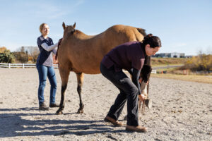
On palpation, clinicians note any muscle spasms, tightness, fibrosis, and pain reactions—all indicators of muscular dysfunction. This hands-on exam also includes mobilization of the neck and back to evaluate range of motion.
Next up is the dynamic exam. The veterinarian, using his or her trained eye, looks for areas of stiffness and abnormality in the horse moving at all three gaits, ideally on different surfaces.
The final step is diagnostic imaging.
- Radiography allows practitioners to evaluate conformation and alignment of vertebrae, as well as diagnose spinal stenosis (narrowing), osteoarthritis, malformation, or fractures.
- Myography helps veterinarians further investigate suspected cases of stenosis.
- Ultrasonography remains the top choice for diagnosing both soft tissue injuries and osteoarthritis, said Waxman. Veterinarians can assess the intervertebral discs of the lumbosacral vertebrae and part of the sacroiliac joint via rectal ultrasonography.
- Nuclear scintigraphy is a valuable tool for localizing areas of bone turnover, which is associated with bone inflammation.
All diagnostic modalities have their advantages and limitations, and practitioners often use them in conjunction with one another. Nevertheless, the equine anatomy remains multilayered and complex and can leave even the experts wondering whether a suspicious diagnostic image is linked to a clinically significant injury. “Fortunately, horses have at least two of most things,” Waxman noted. “When in doubt, use the opposing structure to compare questionable anatomy.”
See “How to Treat Neck and Back Problems in The Horse” for the continuation of this series.