The suspensory ligament is a critical component of the equine athlete’s musculoskeletal apparatus. It originates at the back of the horse’s cannon bone in both the fore- and hind limbs, just beneath the carpus (knee) and tarsus (hock) joints, respectively, and travels down the limb before splitting above the fetlock and inserting onto the sesamoid bones. Its main job is to support the fetlock joint, particularly during exercise.
As a result of repetitive loading during work, subtle damage to ligament fibers can occur and accumulate over time. So, what happens when this important structure gets injured? In this case study we will follow a suspensory ligament injury in a young Quarter Horse mare named Happy, diagnosed and treated at Colorado State University, where I am a resident in equine diagnostic imaging.
Pinpointing the Problem
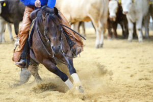
Happy is a 3-year-old reining mount. Her owners brought her to equine sports medicine clinician Katie Seabaugh, DVM, MS, Dipl. ACVSMR, with a complaint she was struggling at the lope, particularly on the left lead. On thorough examination Happy showed a positive response to flexion of the left hind limb, indicating lameness. Seabaugh performed a nerve block injection to localize the source of Happy’s discomfort. A nerve block intended to numb sensation at the origin of the suspensory ligament (also termed the proximal suspensory ligament) yielded a significant improvement in the both the lameness and the quality of the lope. Based on these findings, we performed an ultrasound exam of the proximal suspensory ligament and adjacent structures.
In the hands of experienced operators, ultrasound is a very useful modality to image the soft tissues of the cannon bone region, including the suspensory ligament. Abnormal changes within the proximal suspensory ligament typically include enlargement, where the margins become rounded, with or without dark (hypoechoic) areas of fiber damage/tearing. Often, the surface of the cannon bone where the ligament originates remodels along with the ligament changes and develops a “scooped out” and/or irregular appearance. Images from the ultrasound exam I performed on Happy demonstrate all these features, including a focal tear of the ligament at its interface with the cannon bone. Based on these changes, we diagnosed Happy with a proximal suspensory ligament injury, also referred to as proximal suspensory desmopathy.

Getting Happy Back on Track
Several treatment and management options are available for proximal suspensory desmopathy, and the one your veterinarian chooses likely depends on the exact nature of your horse’s injury. Regardless of the treatments used, the mainstay of recovery is a well-curated rest and rehabilitation program that evolves as the horse heals. This means the length of rehab can vary widely between horses, depending on the degree of injury and their progress through the rehabilitation program. In Happy’s case we immediately initiated a period of stall rest with extremely limited hand-walking. In addition, we administered a series of shock wave treatments. Extracorporeal shock wave therapy involves applying a probe to the skin that emits sound waves that extend into tissues overlying the injured region. The sound waves result in pulses of energy that have been shown to relieve pain associated with the injury as well as stimulate the healing process. We administer multiple treatments, usually three sessions spaced two weeks apart.
Six weeks after diagnosing Happy’s injury, once her course of shock wave therapy was complete, we performed a recheck ultrasound of the suspensory ligament. At this time her lameness was improved; however, the ultrasound picture was almost unchanged from the previous scan. Why would this be, and is it cause for concern? This is, in fact, a relatively common feature of suspensory ligament injuries; the ligament is unlikely to fully remodel such that it returns to its preinjury ultrasonographic appearance. This is important to realize so as to not be discouraged when rechecking these horses.
While we might not expect to see improvement on ultrasound in the early stages, we conduct these rechecks because it is important to ensure the injury has not worsened since the previous scan. This is particularly important if the original ultrasound was performed immediately after the sudden onset of a lameness, because injuries can mature and develop for a period after the initial insult. Therefore, a static ultrasound appearance in a horse with improving lameness is a common and welcome scenario in horses with this type of injury and might prompt a gradual increase in the amount of exercise deemed safe for the horse to perform.
We managed Happy in this way, with lameness examinations at six- to eight-week intervals, gradual increases in the duration and intensity of exercise, and a recommendation to ice the affected region for 20-30 minutes after riding. Happy had several recheck ultrasound exams throughout the healing process (prior to each recommended increase in her workload). The appearance of her suspensory ligament on ultrasound remained unchanged throughout her rehabilitation program. Six months following her original diagnosis, we estimated Happy’s lameness had improved by 85%. At this time she was able to perform walk, jog, and lope work. At the time of writing, we were formulating a plan to reintroduce maneuvers (spins, stops, etc.).
Progress and Prognosis
Happy made good progress in healing with a fairly conservative treatment regimen. There are additional treatment types available for these injuries, which your veterinarian might recommend, depending on the nature of the lesion. In recently injured ligaments it might be possible to inject a biologic product—such as platelet-rich plasma (PRP) or stem cells—directly into regions of fiber damage. Where fiber damage is more long-standing or diffuse or where access to the injured area is more challenging, stem cells might be also be injected into the artery supplying the ligament.
In addition, a surgical procedure known as neurectomy and fasciotomy might be a suitable treatment option in certain cases. This procedure involves severing the nerve that supplies the injured ligament so as to alleviate the pain originating from this region, as well as cutting into the overlying tissues to relieve any local pressure. When considering this surgery, veterinarians might pursue additional imaging of the proximal suspensory region—typically an MRI scan—which will provide more detailed information about the ligament damage and allow them to examine the adjacent cannon bone for associated inflammation or bruising that might factor into the horse’s recovery and the expected outcome.
The prognosis following proximal suspensory ligament desmopathy depends on several factors, including the location of the injury (injuries in the forelimb are associated with a better outcome than those in the hind limb), severity, speed of diagnosis, and athletic goals of the owner/rider. Unfortunately, because the ligament will never return to its preinjured state, it is always at risk of re-injury due to the inherent slight weakness in the previously injured fibers. You can mitigate this risk with a comprehensive rehabilitation plan and gradual reintroduction of work under the careful supervision of your veterinarian.